
Page 14 - IJB-10-4
P. 14
International Journal of Bioprinting PAI for 3D bioprinted constructs
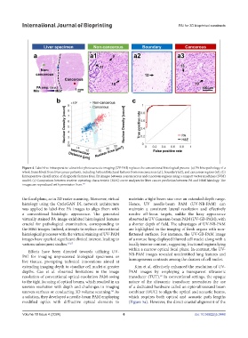
Figure 4. Label-free intraoperative ultraviolet-photoacoustic imaging (UV-PAI) replaces the conventional histological process. (a) PA histopathology of a
whole tissue block from liver cancer patients, including histoarchitectural features from noncancerous (a1), boundary (a2), and cancerous regions (a3). (b)
Intraoperative classification of diagnostic features from PA images between noncancerous and cancerous regions using a support vector machines (SVM)
model. (c) Comparison between receiver operating characteristic (ROC) curve analyses for liver cancer prediction between PA and H&E histology. The
images are reproduced with permission from. 64
the focal plane, as in 2D raster scanning. Moreover, virtual maintain a tight beam size over an extended depth range.
histology using the CycleGAN DL network architecture Hence, UV needle-beam PAM (UV-NB-PAM) can
was applied to label-free PA images to align them with maintain a consistent lateral resolution and effectively
a conventional histologic appearance. The generated resolve off-focus targets, unlike the hazy appearance
virtually stained PA image exhibited histological features observed in UV Gaussian-beam PAM (UV-GB-PAM), with
crucial for pathological examination, corresponding to a shorter depth of field. The advantages of UV-NB-PAM
the H&E images. Indeed, attempts to replace conventional are highlighted in the imaging of fresh organs with non-
histological processes with the virtual staining of UV-PAM flattened surfaces. For instance, the UV-GB-PAM image
images have sparked significant clinical interest, leading to of a mouse lung displayed blurred cell nuclei along with a
various subsequent studies. 65,67 locally intense contrast, suggesting fractional regions lying
Efforts have been directed towards utilizing UV- within a narrow optical focal plane. In contrast, the UV-
PAI for imaging unprocessed biological specimens or NB-PAM images revealed unidentified lung features and
live tissues, prompting technical innovations aimed at homogeneous contrasts among the clusters of cell nuclei.
extending imaging depth to visualize cell nuclei at greater Kim et al. effectively enhanced the resolution of UV-
depths. Cao et al. observed limitations in the image PAM images by employing a transparent ultrasonic
resolution of conventional optical-resolution PAM owing transducer (TUT). In conventional settings, the opaque
69
to the tight focusing of optical beams, which resulted in an nature of the ultrasonic transducer necessitates the use
uneven resolution with depth and challenges in imaging of a dedicated hardware called an opto-ultrasound beam
uneven surfaces or conducting 3D volume scanning. As combiner (OUC) to align the optical and acoustic beams,
68
a solution, they developed a needle-beam PAM employing which requires both optical and acoustic path lengths
modified optics with diffractive optical elements to (Figure 5a). However, the direct coaxial alignment of the
Volume 10 Issue 4 (2024) 6 doi: 10.36922/ijb.3448