
Page 19 - IJB-10-4
P. 19
International Journal of Bioprinting PAI for 3D bioprinted constructs
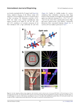
are directly correlated with the PA signal level (Figure 8a). (Figure 8b). Finally, the Achilles tendon of a mouse,
Hence, repetitive imaging of the fibrous tissues with comprising three subtendons connecting three large
variations in the azimuthal axis allows for the identification muscles to the calcaneus, was imaged. The average azimuth
of fiber orientation. The dichroism sensitivity of DS- angles were distinctly measured to be −27.6°, −6.5°, and
PAM was tested using crossed monofilament wire +14.5° for the soleus (Sol), medial gastrocnemius (Mg),
targets scanned at six angles (0°, 30°, 60°, 90°, 120°, and lateral gastrocnemius (Lg) tendons, respectively,
and 150°), resulting in an accurate estimation of the delineating their anatomical positions (Figure 8c). A
fiber orientation with a high uniformity for each wire summary of these studies is provided in Table 1.
Figure 8. Azimuth mapping of fibrous tissue alignment with dichroism-sensitive photoacoustic microscopy (DS-PAM). (a) Principle diagram of linear
dichroism-based PA effect. (b) Validative DS-PAM image of a crossed-wire phantom, demonstrating uniform orientation angle from individual string. (c)
DS-PAM images of a mouse’s Achilles tendon connecting the calcaneus to the soleus (Sol), medial gastrocnemius (Mg), and lateral gastrocnemius (Lg)
muscles, and their corresponding azimuth distributions. The images are reproduced with permission from. 79
Volume 10 Issue 4 (2024) 11 doi: 10.36922/ijb.3448