
Page 22 - IJB-10-4
P. 22
International Journal of Bioprinting PAI for 3D bioprinted constructs
of real skin for the same skin phototype, with strong PA a minimally invasive technique that employs NIR light
signal peaks observed where real and synthetic melanin to destroy cancer cells via hyperthermia. Therefore, it
clustered, allowing for the visualization of the melanin is necessary to develop a multifunctional scaffold with a
distribution (Figure 9b). These findings demonstrate the photothermal conversion ability that can address the risk
utility of PAI for evaluating the functional and structural of local cancer recurrence and achieve tissue repair by
fidelity of 3D-bioprinted biomimetic models to real filling the cavities. In this study, the researchers utilized
biological tissues. 3D-printing methods to fabricate porous scaffolds with
3.2. In vivo monitoring of degradable scaffold with precisely controlled structures for tissue repair and
PA contrast photothermal effects (Figure 10a). Alginate-polydopamine
Degradation monitoring of bioprinted scaffolds in the body (Alg-PDA) was used to fabricate the scaffolds. Alg-PDA,
is a vital aspect of tissue engineering. Luo et al. proposed which is a conjugate of alginate and PDA, is a biocompatible
a novel method using 3D bioprinting as a nonsurgical cell-adhesive material that is widely used in soft tissue
treatment for breast cancer. Currently, surgical removal, engineering and adipose tissue regeneration. Alginate was
83
chemotherapy, and radiotherapy are the primary clinical modified using dopamine to introduce catechol moieties,
approaches for treating breast cancer. However, they are resulting in high biocompatibility and cell adhesion. In
associated with high local recurrence rates and a risk of vivo validation using mice confirmed the stability of the
breast tissue loss. In contrast, photothermal therapy is material, demonstrating a photothermal effect on cancer
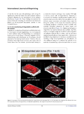
Figure 9. Assessment of human skin phototype phantom using photoacoustic imaging (PAI). (a) 3D-bioprinted skin phantom with different skin tones.
(b) Comparison between PA signals from real skin and mimicked skin. The images are reproduced with permission from. 82
Volume 10 Issue 4 (2024) 14 doi: 10.36922/ijb.3448