
Page 25 - IJB-10-4
P. 25
International Journal of Bioprinting PAI for 3D bioprinted constructs
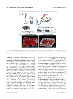
Figure 12. Core-shell hydrogel scaffolds for on-demand drug release in the management of postoperative residual breast cancer. (a) Schematics for printing
process and in vivo tests (b) In vivo PAI of 3D-printed PDA/Alg hollow (with core gels completely released) after implantation for 1 and 7 days. The images
are reproduced with permission from. 85
challenging. Conventional imaging methods, such as X-ray imaging, and the porous structure was visible through the
computed tomography and magnetic resonance imaging, photoacoustic-ultrasound (PA-US) mode, which enabled
have limitations in detecting initial bone tissue formation a clear imaging of even a single printed filament. By
owing to the limited amount of new tissue. To address this comparing the intensity of the PA signal, the researchers
gap, the researchers in this study developed a bimodal could determine which scaffolds could promote faster new
imaging approach that combines NIR fluorescence (NIR- bone formation (Figure 13b). Notably, while ultrasound
FL) and PAI to monitor bone tissue-engineered scaffolds. images suffer from poor in vivo spatial resolution due to
The scaffolds were labeled with a fluorescent probe called tissue interference, PA images are only moderately affected.
turn-on hemicyanine dye (LET-3), which binds to alkaline This study represents a valuable contribution wherein PAI
phosphatase (ALP), a biomarker of bone formation was effectively utilized in combination with other imaging
(Figure 13a). ALP triggered the NIR-FL/PA signal of modalities, such as fluorescence and ultrasound imaging,
LET-3 in scaffolds in vitro and in vivo, allowing for the to noninvasively monitor bone tissue engineering scaffolds
visualization of the osteogenesis-related bioactivity of in real-time during the bone regeneration process.
LET-3-labeled scaffolds. This probe enables researchers
to monitor the early-stage expression of ALP on scaffolds, 3.3. In vivo longitudinal monitoring of angiogenesis
both in vitro and in vivo. Among these techniques, In tissue engineering, numerous studies have utilized PAI
fluorescence imaging is effective in screening the ALP for the nondestructive monitoring of neovascularization
activity in vivo but is suboptimal in generating images to validate the successful in vivo adaptation of functional
of 3D-printed scaffolds with good spatial resolution. In scaffolds implanted in the body. 87–91 For example, Ogunlade
contrast, PAI provides greater details of the scaffolds, et al. observed the longitudinal vascular growth of a
showing a deep penetration depth and high spatial subcutaneously implanted scaffold comprising a vascular
resolution. The scaffold was reconstructed using 3D endothelial growth factor (VEGF)-loaded decellularized
Volume 10 Issue 4 (2024) 17 doi: 10.36922/ijb.3448