
Page 30 - IJB-10-4
P. 30
International Journal of Bioprinting PAI for 3D bioprinted constructs
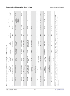
Imaging depth - 3.6 mm 1–5 mm - - - <50 mm <10 mm 26 mm - -
Resolution 4 μm 3.8 μm (OR-PAM) 28 μm (AR-PAM) - - - - - 50–150 μm 113 μm - -
Wavelength 532 nm 532; 590 nm 680–970 nm 808 nm 808 nm 808 nm 710 nm 410–2100 nm 532 nm - 700; 750–850 nm
PA chromophore RBC RBC PDA PDA PDA PDA LET-3 RBC RBC RBC RBC
Imaging target Hydrogel pores Blood vessel, thrombus Synthetic melanin Alg-PDA scaffold PC@ZIF-8@PDA /SerMA hydrogel Alg-PDA scaffold LET-3 labeled PCL/ CS scaffold Blood vessel Blood vessel Blood vessel Blood vessel
Table 2. Summary of the application of photoacoustic imaging (PAI) in 3D bioprinting fields
Printed construct - Multi-patterned printed vessel Skin phantom Scaffold Scaffold Scaffold Scaffold - Hydrogel patch Scaffold Scaffold Abbreviations: Alg, alginate; AR-PAM, acoustic-resolution photoacoustic microscopy; CS, calcium silicate; dECM, decellularized extracellular matrix; GelMA, gelatin methacryloyl; HAMA, methacrylated hyaluronic acid; OR-PAM, optical-resolution photoacoustic microscopy; PCL, polycaprolactone; PDA, polydopamine
Bioprinting material GelMA GelMA, HAMA GelMA Alg-PDA PC@ZIF-8@PDA /SerMA Alg-PDA LET-3 labeled PCL/CS scaffold Tracheal dECM HAMA, Vessel dECM Oxidized hyaluronic acid Hyaluronan-based hydrogel
3D bioprinting application Pore distribution sample Thrombosis phantom Skin phototype phantom Breast cancer photothermal therapy Cartilage reconstruction after chondrosarcoma resection NIR-triggered drug releasing breast implant after lumpectomy Bone regeneration Promoted neovascularization Promoted neovascularization Tissue repair or bone regeneration Cartilage reconstruction
Author Zhao et al. Ma et al. Yim et al. Luo et al. Zhu et al. Wei et al. Yang et al. Ogunlade et al. Hwang et al. Li et al. Tosoratti et al.
Ref. 80 81 82 83 84 85 86 92 93 94 95 modified sericin.
Volume 10 Issue 4 (2024) 22 doi: 10.36922/ijb.3448