
Page 116 - IJB-10-4
P. 116
International Journal of Bioprinting Bioprinting hearing loss treatment
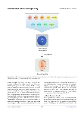
Figure 2. The workflow of auricle defect reconstruction with 3D bioprinting. Abbreviations: ITOP/NIR-DNP: integrated tissue-organ printer/near infrared
-photopolymerization; PCL: polycaprolactone; PGA: poly(glycolic acid).
48
chronic tympanic membrane perforations. In subsequent which demonstrated that various paracrine factors released
research conducted by Reilly et al., it was determined from MSCs, including vascular endothelial growth factor
that bioprinting enhances the stability of TMP healing, (VEGF), transforming growth factor beta 1 (TGF-β1),
demonstrating superior healing properties and feasibility platelet-derived growth factor (PDGF), and insulin-like
as demonstrated in both in vivo and in vitro experiments. growth factor (IGF), play a significant role in promoting
49
In a recent study, Kim et al. investigated the utilization of regeneration of the tympanic membrane. Figure 3 shows
48
growth factors, such as human cord serum (hUCS) and the progression of bioprinting repair in TMP.
basic fibroblast growth factor (bFGF), in conjunction with
bioprinted cell scaffolds, revealing synergistic effects in 2.3. Ossicular discontinuity
promoting the regeneration of the tympanic membrane. Ossiculoplasties play a critical role in addressing persistent
50
The findings indicate that growth factors are essential for conductive hearing loss caused by disruptions in the ossicular
51
facilitating tympanic membrane repair via bioprinting, chain. The utilization of 3D bioprinting, recognized for its
supporting the earlier research conducted by Kim et al. superior plasticity and favorable biocompatibility in both
Volume 10 Issue 4 (2024) 108 doi: 10.36922/ijb.3497