
Page 279 - IJB-10-4
P. 279
International Journal of Bioprinting 3D printing prosthesis for palatal fistula
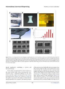
Figure 2. Extraction of airway and bone models and design of speech aid prosthesis. (A) The prosthesis model was assembled with the above models. (B)
The model of the speech aid prosthesis was made, and then (C) the LCD printing machine was used to fabricate the prosthesis. No discernible difference
was observed between the designed models and the finished printed products. (D) The upper airway models before and after repair were imported into the
Ansys 2021 software to divide the CFD model mesh. (E) Almost all the element metrics in mesh quality analysis are above 0.5, indicating high accuracy of
the model. (F) Scanning electron microscopy (SEM) images show a mesh thin scaffold with a pore size of 500×300 microns; scale bars: 200 microns (for
both left and right images).
thereby significantly contributing to precise and of the anterior airway on the affected side was higher than
functional therapy. that on the healthy side due to the invalid cavity caused
by the fistula. We compared the flow velocity of the
In this study, we established five coronal sections on healthy and affected sides during inspiration. The flow
the upper airway model. Upon comparing the cross- velocity of the fistula was much higher than that of other
sectional areas, a significant difference in the areas parts (Figure 3C). The pressure diagram of the restored
before and after repair was observed in the sections upper airway model during inspiration (Figure 3D)
following Section 2 (Figure 3A). As shown in Figure 3B, shows that the pressure on the healthy side and the
the sagittal air pressure on the healthy and affected sides affected side tends to be the same and uniform after the
during inspiration was compared. The negative pressure restoration of the prosthesis. Besides, the large area of
Volume 10 Issue 4 (2024) 271 doi: 10.36922/ijb.2516