
Page 278 - IJB-10-4
P. 278
International Journal of Bioprinting 3D printing prosthesis for palatal fistula
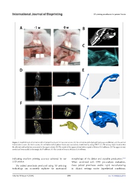
Figure 1. Establishment of animal model of palatal fistula and 3D reconstruction. (A) An animal model of palatal fistula was established, and the animal
healed after a week. (B) After a week, the animal model of palatal fistula was successfully established by using CBCT. (C) The airway mask revealed that
the affected oral cavity was connected to the upper airway. (D) The model of the upper airway before repair in Mimics 21.0 software. (E) The upper airway
model and bone model in Geomagic 2017 software. (F) The model of bone in Mimics 21.0 software.
indicating excellent printing accuracy achieved by our morphology of the defect and expedite production. 39,40
LCD printer. When combined with CFD pre-analysis evaluation,
The palatal prosthesis produced using 3D printing these palatal prostheses enable rapid manufacturing
technology can accurately replicate the anatomical in clinical settings under hypothetical conditions,
Volume 10 Issue 4 (2024) 270 doi: 10.36922/ijb.2516