
Page 273 - IJB-10-4
P. 273
International Journal of Bioprinting 3D printing prosthesis for palatal fistula
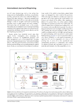
are still some shortcomings, such as low surface free bone model of the rabbit’s airway before palatal fistula
energy and poor hydrophilicity, which may cause friction, repair and designed the initial model of the prosthesis.
and thus, patients may suffer from noticeable allergies to CFD technology was utilized to simulate and analyze the
foreign body after wearing it. Moreover, manufacturers gas flow in the airway, adjusting the model based on the
24
obtain the final products of these commonly used materials pressure and velocity of the airflow. After repairing the
through a series of complex traditional manufacturing defect of the palatal fistula, the airflow pattern exhibits
25
processes, such as mold making and mold pouring. high degree of resemblance to that of the healthy side. The
Information bias will inevitably occur in this process. repaired palatal fistula exhibited good sealing effect, and
Meanwhile, due to the limited types of existing 3D printing the oronasal cavity was effectively separated. Secondly, we
materials for the digital design system, no research has ever designed and synthesized a new light-curable polyurethane
reported that soft elastic materials can be directly used for (PU) printing ink with good biological safety. This ink
the 3D printing of prosthesis. 26,27 could be manufactured by an LCD light-curing 3D printer
Recent studies have identified several areas that for rapid prototyping, and it was utilized to print palatal
require further investigation when evaluating the fistula prosthesis (Scheme 1). In brief, polytetramethylene
different properties of maxillofacial prosthesis and ether glycol (PTMEG) is a soft segment preparation of
their management, such as biocompatibility, cleaning light-cured PU and the preparation of a series of PU
28
29
30
protocols, pigment incorporation, and material bonding printing inks. A series of in vitro experiments were
effects. This study puts forward an optimization method systematically performed to characterize and evaluate
31
for body structure design and a printing strategy for palatal the physicochemical and mechanical properties of the
fistula: Firstly, we reconstructed the mechanical model and materials and determine their biocompatibility in vivo.
Scheme 1. Schematic illustration of the preparation of polyurethane (PU) elastomers, the palatal fistula model design guided by computer fluid dynamics
(CFD) analysis, and the investigations of their performance. (A) Preparation of light-cured PU and configuration of printing ink for palatal prosthesis.
(B) Establishment of an animal model of palatal fistula and prosthesis development using 3D printing based on CFD design. (C) Evaluation of the
properties of light-cured PU elastomers. Abbreviations: CBCT, cone beam computed tomography; HEMA, 2-hydroxyethyl methacrylate; IPDI, isophorone
diisocyanate; PTMEG, polytetramethylene ether glycol.
Volume 10 Issue 4 (2024) 265 doi: 10.36922/ijb.2516