
Page 311 - IJB-10-4
P. 311
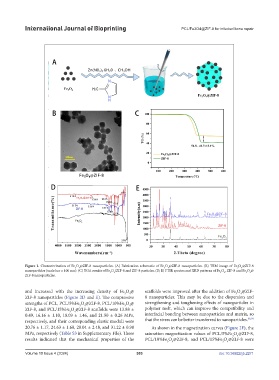
International Journal of Bioprinting                              PCL/Fe3O4@ZIF-8 for infected bone repair


            Figure 1. Characterization of Fe O @ZIF-8 nanoparticles. (A) Fabrication schematic of Fe O @ZIF-8 nanoparticles. (B) TEM image of Fe O @ZIF-8
                                                                         4
                                                                                                      3
                                                                                                        4
                                                                       3
                                   4
                                 3
            nanoparticles (scale bar = 100 nm). (C) TGA results of Fe O /ZIF-8 and ZIF-8 particles. (D, E) FTIR spectra and XRD patterns of Fe O , ZIF-8 and Fe O @
                                                                                               3
                                                                                                 4
                                                  4
                                                3
                                                                                                            4
                                                                                                          3
            ZIF-8 nanoparticles.
            and increased with the increasing density of Fe O @  scaffolds were improved after the addition of Fe O @ZIF-
                                                        4
                                                                                                      3
                                                      3
                                                                                                        4
            ZIF-8 nanoparticles (Figure 2D and E). The compressive   8  nanoparticles.  This  may  be  due  to  the  dispersion  and
            strengths of PCL, PCL/5%Fe O @ZIF-8, PCL/10%Fe O @  strengthening and toughening effects of nanoparticles in
                                                        4
                                   3
                                     4
                                                      3
            ZIF-8, and PCL/15%Fe O @ZIF-8 scaffolds were 13.88 ±   polymer melt, which can improve the compatibility and
                               3
                                 4
            0.49, 16.16 ± 1.10, 18.09 ± 1.46, and 21.50 ± 0.26 MPa,   interfacial bonding between nanoparticles and matrix, so
            respectively, and their corresponding elastic moduli were   that the stress can be better transferred to nanoparticles. 38,39
            20.76 ± 1.17, 24.63 ± 1.60, 28.04 ± 2.48, and 31.22 ± 0.98   As shown in the magnetization curves (Figure 2F), the
            MPa, respectively (Table S3 in Supplementary File). These   saturation magnetization values of PCL/5%Fe O @ZIF-8,
                                                                                                      4
                                                                                                    3
            results indicated that the mechanical properties of the   PCL/10%Fe O @ZIF-8, and PCL/15%Fe O @ZIF-8 were
                                                                        3  4                    3  4
            Volume 10 Issue 4 (2024)                       303                                doi: 10.36922/ijb.2271