
Page 314 - IJB-10-4
P. 314
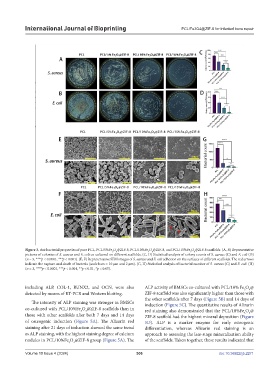
International Journal of Bioprinting                              PCL/Fe3O4@ZIF-8 for infected bone repair


            Figure 3. Antibacterial properties of pure PCL, PCL/5%Fe O @ZIF-8, PCL/10%Fe O @ZIF-8, and PCL/15%Fe O @ZIF-8 scaffolds. (A, B) Representative
                                                 3
                                                   4
                                                                                    4
                                                                  4
                                                                                  3
                                                                3
            pictures of colonies of S. aureus and E. coli co-cultured on different scaffolds. (C, D) Statistical analysis of colony counts of S. aureus (C) and E. coli (D)
            (n = 3, ****p < 0.0001, ***p < 0.001). (E, F) Representative SEM images of S. aureus and E. coli adhesion on the surfaces of different scaffolds. The red arrows
            indicate the rupture and death of bacteria (scale bars = 10 µm and 2 µm). (G, H) Statistical analysis of bacterial number of S. aureus (G) and E. coli. (H)
            (n = 3, ****p < 0.0001, ***p < 0.001, **p < 0.01, *p < 0.05).
            including ALP, COL-1, RUNX2, and OCN, were also    ALP activity of BMSCs co-cultured with PCL/10% Fe O @
                                                                                                           4
                                                                                                         3
            detected by means of RT-PCR and Western blotting.  ZIF-8 scaffold was also significantly higher than those with
                                                               the other scaffolds after 7 days (Figure 5B) and 14 days of
               The intensity of ALP staining was stronger in BMSCs   induction (Figure 5C). The quantitative results of Alizarin
            co-cultured with PCL/10%Fe O @ZIF-8 scaffolds than in   red staining also demonstrated that the PCL/10%Fe O @
                                   3
                                     4
                                                                                                         3
                                                                                                           4
            those with other scaffolds after both 7 days and 14 days   ZIF-8 scaffold had the highest mineral deposition (Figure
            of osteogenic induction (Figure 5A). The Alizarin red   5D). ALP is a marker enzyme for early osteogenic
            staining after 21 days of induction showed the same trend   differentiation, whereas Alizarin red staining is an
            as ALP staining, with the highest staining degree of calcium   approach to assessing the late-stage mineralization ability
            nodules in PCL/10%Fe O @ZIF-8 group (Figure 5A). The   of the scaffolds. Taken together, these results indicated that
                              3  4
            Volume 10 Issue 4 (2024)                       306                                doi: 10.36922/ijb.2271