
Page 317 - IJB-10-4
P. 317
International Journal of Bioprinting PCL/Fe3O4@ZIF-8 for infected bone repair
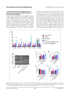
3.6. Wnt/β-catenin pathway participating in the of Dkk1, the gene expressions of β-catenin, Runx2, C-myc,
induction of osteogenesis in BMSCs treated with Gsk-3β, C-jun, and Tcf-7 were evaluated by RT-PCR, and
PCL/Fe O @ZIF-8 scaffolds the protein expressions of β-catenin, RUNX2, GSK-3β,
4
3
To explore the role of the Wnt/β-catenin signaling pathway and p-GSK-3β were evaluated by Western blotting. The
in the osteogenic differentiation of BMSCs co-cultured expression levels of β-catenin, Runx2, C-myc, C-jun, and
with the scaffolds, BMSCs were divided into control group, Tcf-7 were significantly higher in BMSCs co-cultured with
control + Dickkopf-related protein 1 (DKK1, a typical PCL/10% Fe O @ZIF-8 scaffolds than those in the control
3
4
inhibitor of the Wnt/β-catenin pathway) group, PCL group and PCL group, while the expression level of Gsk-
29
group, PCL + DKK1 group, PCL/10%Fe O @ZIF-8 group, 3β was significantly lower (Figure 6A). These effects were
4
3
and PCL/10%Fe O @ZIF-8 + DKK1 group. After BMSCs obviously reversed by the addition of DKK1. The protein
4
3
were co-cultured with different scaffolds in osteogenic expression levels of β-catenin, RUNX2, GSK-3β, and
induction medium for 7 days, with or without 100 ng/mL p-GSK-3β were significantly higher in the PCL/10%Fe O @
3 4
Figure 6. Wnt/β-catenin pathway in BMSCs co-cultured with PCL/Fe O @ZIF-8 scaffolds. (A) Gene expression of β-catenin, Runx2, Tcf-7, C-jun, C-myc,
4
3
and Gsk-3β in the Wnt/β-catenin pathway (n = 3, ***p < 0.001, **p < 0.01, *p < 0.05). (B) Protein expression of β-catenin, RUNX2, GSK-3β, and P-GSK-3β.
(C–F) Semi-quantitative analysis of protein expression levels through ImageJ software (n = 3, ***p < 0.001, **p < 0.01, *p < 0.05).
Volume 10 Issue 4 (2024) 309 doi: 10.36922/ijb.2271