
Page 320 - IJB-10-4
P. 320
International Journal of Bioprinting PCL/Fe3O4@ZIF-8 for infected bone repair
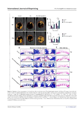
Figure 8. Evaluation of bone regeneration in infected bone defect in vivo by micro-CT and histological analysis. (A) Micro-CT images of rat skulls after
6 and 12 weeks of scaffolds implantation; the red circles show the initial size of cranial defect, and the green arrows indicate marginal bone resorption
(scale bar = 1 mm). (B, C) Quantitative analysis of micro-CT, including BV/TV and Tb.N (n = 3, ****p < 0.0001, ***p < 0.001, **p < 0.01, *p < 0.05).
(D) Masson’s trichrome staining of bone regeneration in rat skulls; magnified images in left black boxes of the first column are displayed in the second
column, and those in the right black boxes are displayed in the third column (scale bars = 1 mm and 200 µm). (E) H&E staining of bone regeneration at
the same sites of rat skulls as stained by Masson’s trichrome staining (scale bar = 1 mm). Abbreviations: HB, host bone; Nb, new bone; MS, residual scaffold
material; FCT, fibrous connective tissue.
Volume 10 Issue 4 (2024) 312 doi: 10.36922/ijb.2271