
Page 350 - IJB-10-4
P. 350
International Journal of Bioprinting Stiffness of scaffold-mediated immune response
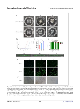
Figure 2. Printability and biocompatibility of macrophage-laden bioinks with different stiffness. (A) Macroscopic imaging of bioprinted and crosslinked
macrophage-laden scaffolds S1, S2, and S3. (B) Degradation of macrophage-laden scaffolds S1, S2, and S3 immersed in PBS for 60 h in vitro. (C) Swelling
rate of macrophage-laden scaffolds S1, S2, and S3 immersed in PBS for 24 h in vitro (n = 3, ****P < 0.0001); (D) Representative 3D imaging of macrophage-
laden scaffolds S1, S2, and S3 after printing; scale bar = 200 μm. (E) Representative imaging of Live/Dead staining with macrophage-laden scaffolds S1,
S2, and S3 at day 1 and day 7; viewed with 50× magnification; scale bar = 500 μm. (F) Microscopic imaging of macrophage-laden scaffolds S1, S2, and
S3 at day 7; viewed with 50× magnification; scale bar = 500 μm. (G) Quantitative analysis of live and dead cell at day 1 and day 7 by two-way ANOVA
(n = 3; *P < 0.05; **P < 0.01; ns: no significance). Notes: S1, low stiffness; S2, medium stiffness, S3: high stiffness.
Volume 10 Issue 4 (2024) 342 doi: 10.36922/ijb.2874