
Page 353 - IJB-10-4
P. 353
International Journal of Bioprinting Stiffness of scaffold-mediated immune response
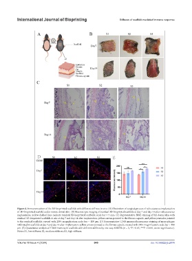
Figure 4. Immunoreaction of the 3D-bioprinted scaffolds with different stiffness in vivo. (A) Illustration of surgical process of subcutaneous implantation
of 3D-bioprinted scaffold under mouse dorsal skin. (B) Macroscopic imaging of residual 3D-bioprinted scaffolds at day 7 and day 14 after subcutaneous
implantation; yellow dashed lines indicate residual 3D-bioprinted scaffolds; scale bar = 5 mm. (C) Representative H&E staining of full dorsal skin with
residual 3D-bioprinted scaffolds in situ at day 7 and day 14 after implantation; yellow arrows pointed to the fibrous capsule, and yellow pentacles pointed
to the residual scaffolds; viewed with 200× magnification; scale bar = 100 μm. (D) Representative CD68 immunofluorescence staining of macrophages
infiltrated in scaffolds at day 7 and day 14 after implantation; yellow arrows pointed to the fibrous capsule; viewed with 200× magnification; scale bar = 100
μm. (E) Quantitative analysis of CD68 intensity in scaffolds with different stiffness by one-way ANOVA (n = 3, *P < 0.05, ***P < 0.001, ns: no significance).
Notes: S1, low stiffness; S2, medium stiffness; S3, high stiffness.
Volume 10 Issue 4 (2024) 345 doi: 10.36922/ijb.2874