
Page 352 - IJB-10-4
P. 352
International Journal of Bioprinting Stiffness of scaffold-mediated immune response
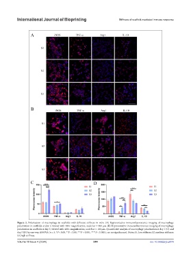
Figure 3. Polarization of macrophage in scaffolds with different stiffness in vitro. (A) Representative immunofluorescence imaging of macrophage
polarization in scaffolds at day 1; viewed with 400× magnification; scale bar = 100 μm. (B) Representative immunofluorescence imaging of macrophage
polarization in scaffolds at day 3; viewed with 400× magnification; scale bar = 100 μm. Quantitative analysis of macrophage polarization at day 1 (C) and
day 3 (D) by one-way ANOVA (n = 3, *P < 0.05, **P < 0.01, ***P < 0.001, ****P < 0.0001, ns: no significance). Notes: S1, low stiffness; S2, medium stiffness;
S3, high stiffness.
Volume 10 Issue 4 (2024) 344 doi: 10.36922/ijb.2874