
Page 43 - IJB-10-4
P. 43
International Journal of Bioprinting 3D bioprinting in otorhinolaryngology
3D structures demonstrated higher cellular activity structures with dECM, and the structures displayed
(supported by the genetic profile), which was conducive improved mechanical stability in cell cultures in vitro.
to tissue differentiation and growth (Figure 3B). In Furthermore, cell differentiation was observed as mouse
26
another study, a 3D scaffold was fabricated using tempo- fibroblasts were converted into myofibroblasts, indicating
oxidized cellulose nanofiber (TOCN), dECM, and SA, tissue repair. The findings of this study could be replicated
89
and researchers evaluated the properties of the scaffolds for repairing tubular organs, i.e., trachea and esophagus.
at different ratios of TOCN and dECM. The results The major challenge of this approach in
indicated that high ratios of dECM in scaffolds provided otorhinolaryngology is the tedious and difficult extraction
a favorable microenvironment for cell proliferation and of dECM from the small organs (e.g., vocal cord). Brown
promoted chondrogenesis via upregulated expression et al. extracted dECM from the vocal cords and small
of cartilage-specific markers. Yeleswarapu et al. used intestinal submucosa tissues and revealed that both
88
stereolithography (SLA) to print self-supporting tubular had relatively similar proteomic characteristics and
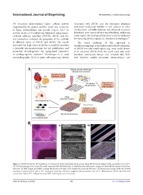
Figure 3. DECM bioinks for 3D bioprinting. (A) Schematic of the tissue bioprinting process using dECM bioink (adapted with permission from ref. ).
26
(B) The bioprinting process of specific tissue constructs with dECM bioinks. (i) Representative microscopic images of a heart dECM construct (scale bar,
400 mm). (ii) SEM images of a hybrid cartilage dECM structure with a PCL framework (scale bar, 400 mm). (iii) Microscopic images of the cell-printed
structure of adipose dECM with a PCL framework (scale bar, 400 mm) (adapted with permission from ref. ). Abbreviations: dECM: decellularized
26
extracellular matrix; PCL: Polycaprolactone; SEM: Scanning electron microscopy.
Volume 10 Issue 4 (2024) 35 doi: 10.36922/ijb.3006