
Page 40 - IJB-10-4
P. 40
International Journal of Bioprinting 3D bioprinting in otorhinolaryngology
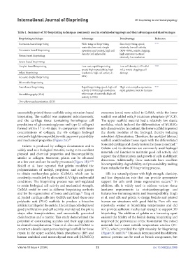
Table 1. Summary of 3D bioprinting techniques commonly used in otorhinolaryngology and their advantages and disadvantages
Bioprinting technique Advantage Disadvantage Reference
Extrusion-based bioprinting Wide range of bioprinting Slow bioprinting speed; 23-32
materials; low cost; simple relatively low cell activity
Pneumatic-based bioprinting operation and control; high cell (40%–80%); nozzle clogging;
density (cell spheroids) high exposure to stress;
Piston-based bioprinting relatively low resolution
Screw-based bioprinting
6
Droplet-based bioprinting Low cost; rapid bioprinting Low cell density (<10 cells/ 33-44
speed; high repeatability; high mL); nozzle clogging; cell
Inkjet bioprinting resolution; high cell activity (> damage
Acoustic droplet bioprinting 85%)
Microvalve bioprinting
Laser-based bioprinting Rapid bioprinting speed; high cell High cost; complex operation; 45-56
activity (>95%); high resolution; rapid gelation; toxic to humans
Stereolithography (SLA) wide range of materials; high cell
activity (>95%)
Two-photon polymerization (2PP)
successfully printed tissue scaffolds using extrusion-based exosomes (exos) were added to GelMA, while the lower
bioprinting. The scaffold was implanted subcutaneously, scaffold was added with β-tricalcium phosphate (β-TCP).
and the cartilage tissue (containing homologous cell The upper scaffold material had a relatively low elastic
populations of glycosaminoglycans and type II collagen) modulus, which induced the differentiation of hUMSCs
formed within 17 to 40 days. In comparison with lower into chondrocytes. In contrast, the lower scaffold improved
concentrations of collagen, the 4% collagen hydrogel the elastic modulus of the hydrogel, thereby inducing
retained a high biocompatibility with improved printability osteoblast differentiation. Therefore, the modified bilateral
and mechanical properties (Figure 2A). 68 scaffold could mediate tissue repair with the differentiated
bone and cartilage and closely restore the tissue to normal.
73
Gelatin is produced by collagen denaturation and is
widely used as a biological material, owing to its excellent Gelatin and its derivatives are commonly used hydrogel
physical and chemical properties and biocompatibility, materials and function to maintain good cell activity and
similar to collagen. Moreover, gelatin can be obtained support the differentiation and growth of cells in different
at a low cost and can be easily processed (Figure 2B). 69,70 directions. Additionally, these materials have excellent
Bedell et al. have reported that gelatin modified the biocompatibility, degradability, and processability, making
polymerization of methyl, propylene, and acyl groups them valuable for the 3D bioprinting process.
to obtain methacrylate gelatin (GelMA), which can be Silk is a natural polymer with high strength, elasticity,
covalently crosslinked by ultraviolet (UV) light under mild and low degradation rate that can provide appropriate
conditions. The bioprinting process was well-regulated support for cells until tissue regeneration occurs. In
74
to retain biological cell activity and mechanical strength. addition, silk is widely used to address various tissue
GelMA could be used in different bioprinting methods hardness requirements in otorhinolaryngology and
and for the regeneration of bones and cartilage. Tang et features low immunogenicity and biocompatibility. Singh
71
al. loaded cartilage cells into GelMA and fixed them onto et al. used silk and gelatin to design bioinks and printed
polylactic acid (PLA) scaffolds to produce a bioactive human ear structures with good fidelity. Pure silk was
artificial cartilage for the auricle. The cartilage cells displayed reportedly weaker at bioprinting temperatures and did
good proliferation and cell activity, maintained the pinna not provide sufficient mechanical strength to support 3D
shape after transplantation, and successfully generated bioprinting. The addition of gelatin as a leavening agent
chondrocytes and a matrix. This study demonstrated the ensured the fidelity of the bioink during bioprinting and
potential of constructing auricles and their applications improved the performance of the biomaterial. The mixed
in clinical transplantation. Sun et al. also used GelMA to materials had a more suitable temperature window (25–
72
construct a double-layer porous hydrogel scaffold for tissue 35°C), which provided the right viscosity for bioprinting
repair. In the upper scaffold, black phosphorus (BP) and (Figure 2C and D). This study demonstrated that different
75
human umbilical cord mesenchymal stem cell (hUMSCs) natural proteins can be used as bioink components, but
Volume 10 Issue 4 (2024) 32 doi: 10.36922/ijb.3006