
Page 49 - IJB-10-4
P. 49
International Journal of Bioprinting 3D bioprinting in otorhinolaryngology
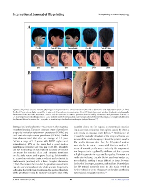
Figure 5. 3D-printed ears and implants. (A) Images of the patient before ear reconstruction (Pre-OP) to 30 months post-implantation (Post-OP 30m).
The Pre-OP microtic ear had a peanut-like structure. At 30 months post-reconstruction (Post-OP 30m), the reconstructed ear displayed typical auricular
137
features with helix, anti-helix, and cavum conchae, and the reconstructed ear was symmetrical to the healthy ear (adapted with permission from ref. ).
(B) A cartilage framework (designed based on the patient’s unaffected contralateral ear) was implanted into the suprafascial plane of a right radial forearm
139
free flap and allowed to mature for 1 year prior to transferring to the head and neck region (adapted from ref. ).
damaged and artificial ossicle replacement is often required ossicular chain. In this regard, a customized ossicular
to restore hearing. The most common types of prostheses chain can treat conductive hearing loss caused by chronic
are partial ossicular replacement prostheses (PORPs) and otitis media or ossicular chain defects. Heikkinen et al.
140
total ossicular replacement prostheses (TORPs). Studies printed the ossicular structure of the temporal bones and
have demonstrated that after an average of 2.5 years measured the conduction properties of the printed ossicles.
(within a range of 1–7 years) with TORP replacement, The results demonstrated that the 3D-printed ossicles
approximately 49% of the cases had a good positive were similar to mature commercial titanium ossicles in
audiological outcome (air-bone gap ≤ 20 dB). Therefore, terms of acoustic performance, whereby the response at
the 3D bioprinting of personalized ossicular prostheses low frequencies is regulated by stiffness and the response
can repair the ossicular chain and tympanic membrane
to effectively restore and improve hearing. Sokołowski et at high frequencies is regulated by quality. However, the
al. printed an ossicular chain prosthesis and evaluated its study also indicated that the bioink used was harder and
performance (motion) with a laser Doppler vibrometer more flexible, making it more difficult to insert between
(LDV). The motion threshold of the prosthesis was close to the head of the stapes, eardrum, and malleus. Nonetheless,
that of a conventional ossicular chain at some frequencies, the 3D-printed materials used in this study could be
indicating that with its customization, the motion threshold further refined in terms of accuracy to develop an effective
of the prosthesis would be relatively similar to that of the personalized ossicular construct. 141
Volume 10 Issue 4 (2024) 41 doi: 10.36922/ijb.3006