
Page 51 - IJB-10-4
P. 51
International Journal of Bioprinting 3D bioprinting in otorhinolaryngology
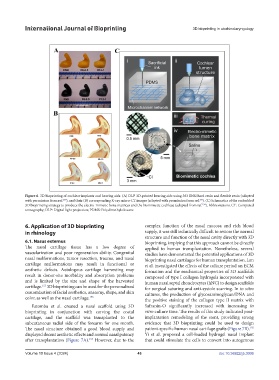
Figure 6. 3D bioprinting of cochlear implants and hearing aids. (A) DLP 3D-printed hearing aids using 363 ENG hard resin and flexible resin (adapted
150
150
with permission from ref. ), and their (B) corresponding X-ray micro-CT images (adapted with permission from ref. ). (C) Schematics of the embedded
154
3D bioprinting strategy to produce the electro-mimetic bone matrices and the biomimetic cochleae (adapted from ref. ). Abbreviations: CT: Computed
tomography; DLP: Digital light projection; PDMS: Polydimethylsiloxane.
6. Application of 3D bioprinting complex function of the nasal mucosa and rich blood
in rhinology supply, it was still technically difficult to restore the normal
structure and function of the nasal cavity directly with 3D
6.1. Nasus externus bioprinting, implying that this approach cannot be directly
The nasal cartilage tissue has a low degree of applied to human transplantation. Nonetheless, several
vascularization and poor regeneration ability. Congenital studies have demonstrated the potential applications of 3D
nasal malformations, tumor resection, trauma, and nasal bioprinting nasal cartilages for human transplantation. Lan
cartilage malformations may result in functional or et al. investigated the effects of the culture period on ECM
aesthetic defects. Autologous cartilage harvesting may formation and the mechanical properties of 3D scaffolds
result in donor-site morbidity and absorption problems composed of type I collagen hydrogels incorporated with
and is limited by the size and shape of the harvested human nasal septal chondrocytes (hNC) to design scaffolds
cartilage. 3D bioprinting can be used for the personalized for surgical suturing and anti-systole scarring. In in vitro
155
customization of facial aesthetics, anatomy, shape, and skin cultures, the production of glycosaminoglycan/DNA and
color, as well as the nasal cartilage. the positive staining of the collagen type II matrix with
156
Estomba et al. created a nasal scaffold using 3D Safranin-O significantly increased with increasing in
bioprinting in conjunction with carving the costal vitro culture time. The results of this study indicated post-
cartilage, and the scaffold was transplanted to the implantation remodeling of the stent, providing strong
subcutaneous radial side of the forearm for one month. evidence that 3D bioprinting could be used to design
The nasal structure obtained a good blood supply and patient-specific human nasal cartilage grafts (Figure 7B).
155
displayed decent aesthetic effects and normal nasal patency Yi et al. proposed a cell-loaded hydrogel nasal implant
after transplantation (Figure 7A). However, due to the that could stimulate the cells to convert into autogenous
157
Volume 10 Issue 4 (2024) 43 doi: 10.36922/ijb.3006