
Page 56 - IJB-10-4
P. 56
International Journal of Bioprinting 3D bioprinting in otorhinolaryngology
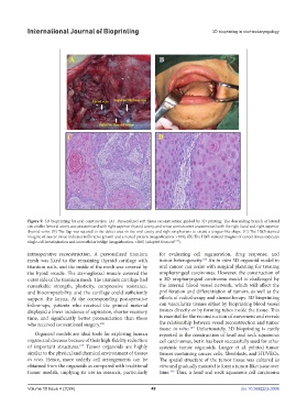
Figure 9. 3D bioprinting for oral construction. (A) Personalized soft tissue reconstruction guided by 3D printing. The descending branch of lateral
circumflex femoral artery was anastomosed with right superior thyroid artery, and venae comites were anastomosed with the right facial and right superior
thyroid veins. (B) The flap was sutured to the defect area on the oral cavity and right oropharynx to create a tongue-like shape. (C) The H&E-stained
imagine of cancer tissue indicates infiltrative growth and a nested pattern (magnification: ×100); (D) The H&E-stained imagine of cancer tissue indicates
179
single-cell keratinization and intercellular bridge (magnification: ×200) (adapted from ref. ).
intraoperative reconstruction. A personalized titanium for evaluating cell regeneration, drug response, and
mesh was fixed to the remaining thyroid cartilage with tumor heterogeneity. An in vitro 3D organoid model in
188
titanium nails, and the inside of the mesh was covered by oral cancer can assist with surgical planning for treating
the hyoid muscle. The sternoglossal muscle covered the oropharyngeal carcinomas. However, the construction of
outer side of the titanium mesh. The titanium cartilage had a 3D oropharyngeal carcinoma model is challenged by
remarkable strength, plasticity, compressive resistance, the internal blood vessel network, which will affect the
and biocompatibility, and the cartilage could sufficiently proliferation and differentiation of tumors, as well as the
support the larynx. At the corresponding postoperative effects of radiotherapy and chemotherapy. 3D bioprinting
follow-ups, patients who received the printed material can vascularize tissues either by bioprinting blood vessel
displayed a lower incidence of aspiration, shorter recovery tissues directly or by forming tubes inside the tissue. This
time, and significantly better pronunciation than those is essential for the reconstruction of movement and reveals
who received conventional surgery. 186 the relationship between vessel reconstruction and tumor
tissue in vitro. Unfortunately, 3D bioprinting is rarely
189
Organoid models are ideal tools for exploring human reported in the construction of head and neck squamous
organs and diseases because of their high-fidelity reduction cell carcinomas, but it has been successfully used for other
of important structures. Tumor organoids are highly systemic tumor organoids. Langer et al. printed tumor
187
similar to the physical and chemical environment of tissues tissues containing cancer cells, fibroblasts, and HUVECs.
in vivo. Hence, more orderly cell arrangements can be The spatial structure of the tumor tissue was cultured in
obtained from the organoids as compared with traditional vitro and gradually matured to form a tumor-like tissue over
tumor models, implying its use in research, particularly time. Thus, a head and neck squamous cell carcinoma
190
Volume 10 Issue 4 (2024) 48 doi: 10.36922/ijb.3006