
Page 52 - IJB-10-4
P. 52
International Journal of Bioprinting 3D bioprinting in otorhinolaryngology
cartilage-like tissues. Although this technique has not anastomosis without scaffold slip-offs by 3D bioprinting a
158
been applied to actual human transplantation, related personalized customized nasal scaffold using Dental SG resin
studies have highlighted the feasibility of the method. cartridges as the material. Similar to this nasal scaffold,
159
Other applications of 3D bioprinting in nasal Jung et al. performed nasal cavity reconstruction surgery for
reconstructions have been reported clinically. A nasal children with congenital nasal deformities. After surgery, a
deformity correction should exhibit and support the shape silicone nasal scaffold was 3D bioprinted and implanted to
of the original structure, e.g., the lip after cleft lip repair. help maintain the shape of the nasal cavity. Three years after
Conventional supports are made using a plaster model and the removal of the nasal scaffolds, the patients’ respiratory
have the disadvantages of high production cost and the function, nasal passage structure, and external nasal shape
inability to further adjust the support position in the future. remained unchanged, demonstrating the feasibility of this
Luo et al. displayed personalized adjustment and good technique (Figure 7C). 160
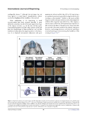
Figure 7. 3D-printed constructs for nasal surgery. (A) The shape and curvature of a 3D anatomical construct were designed based on the patient’s nasal
measurements and anatomy (adapted from ref. ). (B) Gross morphology of nasal constructs and scaffolds before and after implantation. Compared with
157
non-pre-cultured engineered cartilages (empty scaffolds), pre-cultured cartilages were smooth and opaque (adapted with permission from ref. ). (C)
155
A designed stent model for bioprinted nasal construction and the CT image(s) of the patient’s nasal region after construct implantation. (Adapted with
permission from ref. ) The yellow box depicted stent insertion. Abbreviation: CT: Computed tomography.
160
Volume 10 Issue 4 (2024) 44 doi: 10.36922/ijb.3006