
Page 495 - IJB-10-4
P. 495
International Journal of Bioprinting Embedded bioprinting of cartilage
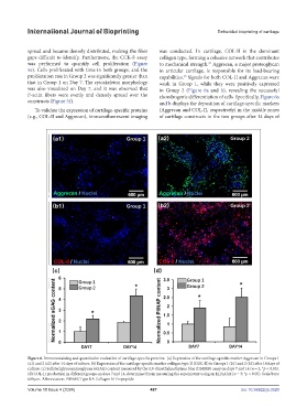
spread and became densely distributed, making the fiber was conducted. In cartilage, COL-II is the dominant
gaps difficult to identify. Furthermore, the CCK-8 assay collagen type, forming a cohesive network that contributes
was performed to quantify cell proliferation (Figure to mechanical strength. Aggrecan, a major proteoglycan
32
5e). Cells proliferated with time in both groups, and the in articular cartilage, is responsible for its load-bearing
proliferation rate in Group 2 was significantly greater than capabilities. Signals for both COL-II and Aggrecan were
33
that in Group 1 on Day 7. The cytoskeleton morphology weak in Group 1, while they were positively expressed
was also visualized on Day 7, and it was observed that in Group 2 (Figure 6a and b), revealing the successful
F-actin fibers were evenly and densely spread over the chondrogenic differentiation of cells. Specifically, Figure 6a
constructs (Figure 5f). and b displays the deposition of cartilage-specific markers
To validate the expression of cartilage-specific proteins (Aggrecan and COL-II, respectively) in the middle zones
(e.g., COL-II and Aggrecan), immunofluorescent imaging of cartilage constructs in the two groups after 14 days of
Figure 6. Immunostaining and quantitative evaluation of cartilage-specific proteins. (a) Expression of the cartilage-specific marker Aggrecan in Groups 1
(a1) and 2 (a2) after 14 days of culture. (b) Expression of the cartilage-specific marker collagen type II (COL-II) in Groups 1 (b1) and 2 (b2) after 14 days of
culture. (c) Sulfated glycosaminoglycan (sGAG) content measured by the 1,9-dimethylmethylene blue (DMMB) assay on days 7 and 14 (n = 3; *p < 0.05).
(d) COL-II production in different groups on days 7 and 14, determined from measuring the supernatant using an ELISA kit (n = 3; *p < 0.05). Scale bars:
600 µm. Abbreviation: PIINAP, Type IIA Collagen N-Propeptide
Volume 10 Issue 4 (2024) 487 doi: 10.36922/ijb.3520