
Page 117 - IJB-5-1
P. 117
Optimization of a 3D-bioprinting process using ultrashort peptide bioinks
thermal waves, generated by air bubbles or piezoelectric Aiming to benefit from the biological properties of the
actuators, which are used for dispensation [3,5] . ultrashort peptides and to combat its unstable mechanical
Extrusion-based bioprinting involves a linear moving properties, we propose introducing a vacuum system
extruder and stage unit which moves across the X-Y-Z into the 3D bioprinting process. The vacuum system,
axes. Bioinks are extruded through nozzles using placed under the print bed, will allow the excess water to
microfluidic pumps, pneumatic pressure, or solenoid be drained and leave the refined structure intact. Aspect
control . biosystems have implemented a similar technology in
[5]
[5]
Laser-assisted bioprinting uses laser beams to print at a their RX1 Bioprinter . This paper will assess the effect
cell resolution . It is becoming widely popular due to its of a vacuum mechanism in optimizing the robotic 3D
[5]
high precision. An additional advantage is that it does not bioprinter to achieve better printing results.
[3]
require a nozzle, which eliminates issues of clogging .
However, for complex structures, recent research indicates 2. Materials and Methods
the advantages in robotic 3D printing as compared to The components of the bioprinter system include the 3D
linear printing. This approach allows for a minimum of bioprinting robotic arm, our custom-designed coaxial
six degrees of freedom, providing much more precision, nozzle, three syringe pumps, and the vacuum mechanism.
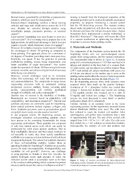
flexibility, and speed. It has the potential to provide The experimental setup is shown in Figure 1a. A vacuum
scaffold-free printing, precise tissue dispensation, and pump with a maximum pressure of −0.35 bar was fitted with
better scalability of organ fabrication . Our system tubings and attached to the hose barb of a vacuum flask.
[6]
adopts the approach of robotic 3D bioprinting to be more A 5-mm suction cup was placed on a rubber stopper. Then,
compact, versatile, and achieve a higher level of accuracy a PET track-etched cell culture membrane with a pore size
while being cost-effective. of 0.4 µm was placed on the suction cup to serve as the
However, several challenges need to be overcome printing surface and to allow the excess of water to penetrate
before this technology will reach full implementation through the membrane and into the flask (Figure 1b).
and commercialization. The complexity to merge tissue For the bioprinting process, three fresh solutions were
engineering processes with an automated printing prepared which later made up the peptide hydrogel.
mechanism involves multiple factors including print A solution of 10 × phosphate buffer was loaded into
quality, vascularization, cell viability, mechanical syringe 1. Serum-free medium was loaded into syringe
strength of scaffolds, and surface topography [1,7] . 2. The peptide powder was weighed out in a ratio of
Another area of concern is the durability of bioinks. 15 mg/mL and loaded into syringe 3. The bioprinting
Biomaterials are assessed based on printability, cell process has been discussed in more detail in another
compatibility, and mechanical properties . Natural and publication (Rauf, 2018, submitted).
[7]
synthetic polymers are commonly used for bioprinting. Cellular viability is an essential factor in the tissue
Some natural polymers include alginate, collagen, and engineering process. In this experiment, neonatal human
fibrin. Synthetic polymers, such as polyethylene glycol dermal fibroblasts (HDFn) were used. The cells were
and poly(L-lactic acid), are also used as bioinks . first cultured to reach the desired cell number. After
[3]
In our proposed robotic 3D bioprinting system, we centrifugation, cell pellets were transferred into a tube
investigate ultrashort self-assembling peptides which of approximately 500 µm. The rate of the seeded cells
7
have proven to be promising biomaterials for tissue ranged from 1.46 × 10 to 1.6 × 10 cells. The cells were
7
engineering applications. These peptides are composed then added into a 1 mL solution of serum-free medium.
of only four natural amino acids which can easily be Subsequently, the mixture of cells and medium was
synthesized by solid phase peptide synthesis. Due to
their amphiphilic character and their innate tendency to
self-assemble in water, they form rapidly nanofibrous
scaffolds in an aqueous solution in forms of soft solid and
transparent hydrogels. The natural but synthetic character
of these self-assembling peptides renders them as
appealing bioinks for bioprinting . Peptides are generally
[8]
known for their biocompatibility, biodegradability, and
suitability for cell growth . However, one challenge
[9]
of using peptides as bioinks is their low viscosity. As
peptide hydrogels retain high amounts of water, the a b
extrusion system tends to accumulate water at the base Figure 1. Setup of vacuum mechanism in three-dimensional
of the construct while printing which weakens the printed bioprinting system. Elements of vacuum mechanism (a) A close up
structure and increases the chance of collapse over time . of peptide printing with vacuum mechanism (b).
[10]
2 International Journal of Bioprinting (2019)–Volume 5, Issue 1