
Page 119 - IJB-5-1
P. 119
Optimization of a 3D-bioprinting process using ultrashort peptide bioinks
G.R.C did microscopic imaging and N. P. supported the
experimental set-up.
Conflicts of Interest
The authors declare that they do not have any competing
interests.
References
a b
1. Chua C, Yeong W. Bioprinting. New Jersey: World Scientific;
st
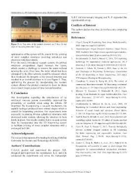
Figure 5. (a) Top view of the printed structure on 1 day, (b) top
view of the same print after 7 days. 2015. https://doi.org/10.1142/9193.
2. Organdonor.gov. Organ Donation Statistics. Organ Donor;
2018. Available from: https://www.organdonor.gov/statistics-
optimization of the system will be crucial for the printing stories/statistics.html. [Last accessed on 2018 Oct 30].
of more complex structures involving curvatures and 3. Sundaramurthi D, Rauf S, Hauser C, 2016, 3D bioprinting
structures with finer details.
Prior the newly introduced vacuum system, the printed technology for regenerative medicine applications. Int J
structures encapsulated liquid between the layers, Bioprint, 2: 9–26. https://doi.org/10.18063/IJB.2016.02.010.
which created a challenge to remove the water without 4. Fermeiro J, Calado M, Correia I, 2015, State of the Art
injuring the print. Over time, the water which had been and Challenges in Bioprinting Technologies, Contribution
entrapped by the fiber network would be released, which of the 3D Bioprinting in Tissue Engineering. 2015 IEEE
then weakened the integrity of the printed structure and 4 Portuguese Meeting on Bioengineering.
th
resulted in an overall decrease in its size Figure 5. Thus,
optimizing the process by incorporating the vacuum 5. Choudhury D, Anand S, Naing M, 2018, The arrival of
system allowed the printed construct to keep its shape commercial bioprinters-towards 3D bioprinting revolution!
over a much longer period of time (several months). Int J Bioprint, 4: 139. https://doi.org/10.18063/ijb.v4i2.139.
6. Mironov V, Kasyanov V, Markwald R, 2011, Organ
5. Conclusion printing: From bioprinter to organ biofabrication line. Curr
Our investigation regarding the introduction of an Opin Biotechnol, 22: 667–673. https://doi.org/10.1016/j.
additional vacuum system successfully improved the copbio.2011.02.006.
printability of scaffold when using the robotic 3D 7. Derakhshanfar S, Mbeleck R, Xu K, et al., 2018, 3D
bioprinter. By incorporating a vacuum mechanism, the bioprinting for biomedical devices and tissue engineering:
peptide hydrogel produced more refined shapes which is
crucial for bioprinting precision. Our experiments were A review of recent trends and advances. Bioact Mater,
successful in printing 40-mm cylindrical structures with 3: 144–156. https://doi.org/10.1016/j.bioactmat.2017.11.008.
decreased water content, allowing the structure to hold 8. Arab W, Rauf S, Al-Harbi O, et al., 2018, Novel ultrashort self-
firmly in place. Our results confirm that a vacuum system assembling peptide bioinks for 3D culture of muscle myoblast
must be incorporated into the 3D bioprinting system cells. Int J Bioprint, 4: 129. https://doi.org/10.18063/ijb.v4i1.129.
to facilitate printing of more complex structures with a 9. Costa R, Rauf S, Hauser C, 2017, Towards biologically
prolonged half-life.
relevant synthetic designer matrices in 3D bioprinting
Acknowledgments for tissue engineering and regenerative medicine. Curr
Opin Biomed Eng, 2: 90–98. https://doi.org/10.1016/j.
The research was supported by funding from King Abdullah
University for Science and Technology (KAUST). cobme.2017.05.001.
10. Hauser C, Deng R, Mishra A, et al., 2011, Natural tri- to
Authors’ Contributions hexapeptides self-assemble in water to amyloid-type fiber
C.A.E.H guided and supervised the project. S.R. aggregates by unexpected-helical intermediate structures.
designed and supervised the experiments. K.K. and Z.K Proc Natl Acad Sci, 108: 1361–1366. https://doi.org/10.1073/
conducted the experiments and wrote the manuscript. pnas.1014796108.
4 International Journal of Bioprinting (2019)–Volume 5, Issue 1