
Page 124 - IJB-5-1
P. 124
Liu F, et al.
A
B
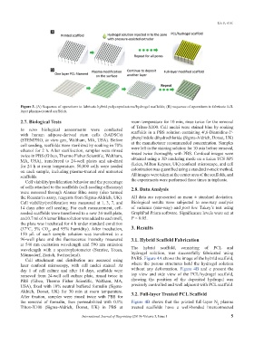
Figure 3. (A) Sequence of operations to fabricate hybrid polycaprolactone/hydrogel scaffolds; (B) sequence of operations to fabricate full-
layer plasma-treated scaffolds.
2.7. Biological Tests room temperature for 10 min, rinse twice for the removal
of Triton-X100. Cell nuclei were stained blue by soaking
In vitro biological assessments were conducted scaffolds in a PBS solution containing 4′,6-Diamidine-2′-
with human adipose-derived stem cells (hADSCs) phenylindole dihydrochloride (Sigma-Aldrich, Dorset, UK)
(STEMPRO, in vitro gen, Waltham, MA, USA). Before
cell seeding, scaffolds were sterilized by soaking in 70% at the manufacturer recommended concentration. Samples
were left in the staining solution for 10 min before removal,
ethanol for 2 h. After sterilization, samples were rinsed
twice in PBS (Gibco, Thermo Fisher Scientific, Waltham, rinsed twice thoroughly with PBS. Confocal images were
MA, USA), transferred to 24-well plates and air-dried obtained using a 3D rendering mode on a Leica TCS SP5
for 24 h at room temperature. 50,000 cells were seeded (Leica, Milton Keynes, UK) confocal microscope, and cell
on each sample, including plasma-treated and untreated colonization was quantified using a standard z-stack method.
scaffolds. All images were taken at the center area of the scaffolds, and
Cell viability/proliferation behavior and the percentage the experiments were performed three times in triplicate.
of cells attached to the scaffolds (cell-seeding efficiency) 2.8. Data Analysis
were assessed through Alamar Blue assay (also termed
the Resazurin assay, reagents from Sigma-Aldrich, UK). All data are represented as mean ± standard deviation.
Cell viability/proliferation was measured at 1, 3, 7, and Biological results were subjected to one-way analysis
14 days after cell seeding. For each measurement, cell- of variance (one-way) and post hoc Tukey’s test using
seeded scaffolds were transferred to a new 24-well plate, GraphPad Prism software. Significance levels were set at
and 0.7 ml of Alamar Blue solution was added to each well, P < 0.05.
the plate was incubated for 4 h under standard condition
(37°C, 5% CO , and 95% humidity). After incubation, 3. Results
2
150 µL of each sample solution was transferred to a
96-well plate and the fluorescence intensity measured 3.1. Hybrid Scaffold Fabrication
at 540 nm excitation wavelength and 590 nm emission
wavelength with a spectrophotometer (Sunrise, Tecan, The hybrid scaffold, consisting of PCL and
Männedorf, Zurich, Switzerland). hydrogel solution, was successfully fabricated using
Cell attachment and distribution are assessed using PABS. Figure 4A shows the image of the hybrid scaffold,
laser confocal microscopy, with cell nuclei stained. At where the porous structures hold the hydrogel solution
day 1 of cell culture and after 14 days, scaffolds were without any deformation. Figure 4B and c present the
removed from 24-well cell culture plate, rinsed twice in top view and side view of the PCL/hydrogel scaffold,
PBS (Gibco, Thermo Fisher Scientific, Waltham, MA, showing the position of the deposited hydrogel was
USA), fixed with 10% neutral buffered formalin (Sigma- precisely controlled and well adjacent with PCL scaffold.
Aldrich, Dorset, UK) for 30 min at room temperature. 3.2. Full-layer Treated PCL Scaffold
After fixation, samples were rinsed twice with PBS for
the removal of formalin, then permeabilized with 0.1% Figure 4B shows that the printed full-layer N plasma
2
Triton-X100 (Sigma-Aldrich, Dorset, UK) in PBS at treated scaffolds have a well-bonded interconnected
International Journal of Bioprinting (2019)–Volume 5, Issue 1 5