
Page 75 - IJB-5-1
P. 75
3D tissue hybrid biofabrication
also misnamed in this context as “biocompatibility),” or first, and then covered by jetted cells [16] . The system
alternatively seeks histo-integration, and therefore the works by printing bioinks in contact mode followed by
implant’s colonization by cells that migrate onto and into its polymerization (e.g., by ultraviolet light) of the printed
structure . As an example, the osteoinductive structures lines, and subsequent jetting of cells onto printed lines
[11]
made of various materials can be considered . This in nanodroplets. One such cycle can be repeated several
[12]
approach is common in regenerative medicine, constituting times resulting in a layered structure . Since hydrogels
[16]
a form of “in vivo tissue engineering” with the host tissue are materials with an absorptive surface, they could take
acting as a bioreactor [13] . In this case, a tissue-repairing up the excess fluid from the jetted droplets.
activity requires merely shape and mechanical function The print and populate method also works with hard
of interest; therefore, the used materials are polymeric or materials, such as polycaprolactone. In tissue engineering,
natural/decellularized fibrillar matrices. Hydrogels, another another version of this method is the colonization with
major medium for tissue engineering, are also occasionally cells of fibrillar and/or porous scaffolds prepared by
injected directly in the recipients to elicit a repairing electrospinning. This is usually performed after the shape
response either by themselves or often as cell carriers . of the scaffold is pre-determined, such as layer and tube .
[14]
[17]
Many bona fide 3D biofabrication tasks primarily target However, for cell placement, simple cell sedimentation
the in vitro applications, and either include the spatial usually does not suffice, because the superficial pores
arrangement of living cells during the printing process or are quickly clogged, preventing their further penetration
make them adhere in a directed manner on specifically in the scaffold. To force them inside of the structure, the
preprinted structures. As a future development, pre- cells may need to be exposed to negative pressure [18] , to
existing structures of living host tissues might be gravitational (centrifugation) or magnetic forces.
[20]
[19]
considered as the equivalent of preprinted structures and
therefore might invite direct in vivo printing approaches 1.1.2 Direct Cells Printing - the Role of Bioinks
onto wound grounds . A typical commercial bioprinter offers the extrusion
[15]
Whatever the approach, biofabrication is done with mode, by which a viscous medium is continuously
the understanding that besides a variety of cells, animal expelled from ready-to-use cartridges through printing
tissues contain various proportions of extracellular matrix needles or nozzles. The extrusion can be achieved through
(ECM) in the form of fibrils, fibrillary networks, sheaths, microvalves propelling the extrudate downward, a plunger
and hydrogels, as well as a fluid phase representing a system, or in the case of very viscous materials, a screw
complex mixture of molecules with metabolic or signaling pump . The cells are delivered in a printing medium, the
[21]
functions. Correspondingly, a re-creation of tissue analogs so-called “bioink,” that contains them during the printing
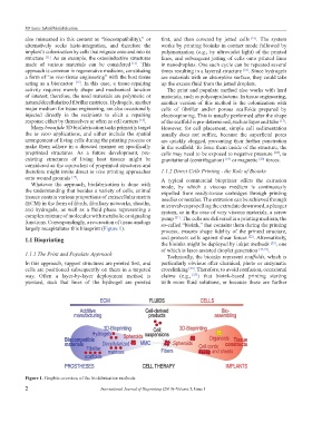
largely recapitulates this blueprint (Figure 1). process, ensures shape fidelity of the printed structure,
[22]
1.1 Bioprinting and protects cells against shear forces . Alternatively,
the bioinks might be deployed by inkjet methods , one
[23]
of which is laser-assisted droplet generation [24,25] .
1.1.1 The Print and Populate Approach
Technically, the bioinks represent scaffolds, which is
In this approach, support structures are printed first, and particularly obvious after chemical, photo or enzymatic
cells are positioned subsequently on them in a targeted crosslinking . Therefore, to avoid confusion, occasional
[26]
way. Often a layer-by-layer deployment method is claims (e.g., ) that bioink-based printing starting
[27]
pursued, such that lines of the hydrogel are printed with more fluid solutions, or because these are further
Figure 1. Graphic overview of the biofabrication methods.
2 International Journal of Bioprinting (2019)–Volume 5, Issue 1