
Page 21 - IJB-10-5
P. 21
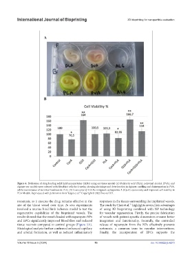
International Journal of Bioprinting                                3D bioprinting for nanoparticle evaluation


            Figure 4. Evaluation of drug loading solid lipid nanoparticles (SLNs) using ear tissue model. (A) Polylactic acid (PLA), polyvinyl alcohol (PVA), and
            alginate ear models were cultured with fibroblast cells for 4 weeks, showing shrinkage and deterioration in alginate, swelling and disintegration in PVA,
            while maintenance of its initial hardness in PLA. (B) Coenzyme Q10-SLNs mitigated cyclosporine A (CycA) cytotoxicity and improved cell viability in
            PLA Models. Reproduced with permission from Yalgın et al.  Copyright © 2022 Elsevier B.V.
                                                  62
            restenosis, as it ensures the drug remains effective at the   responses in the tissues surrounding the implanted vessels.
            site of the blood vessel over time.  In vivo experiments   The study by Choi et al.  highlights several key advantages
                                                                                 71
            involved a murine hind limb ischemia model to test the   of using 3D bioprinting combined with NP  technology
            regenerative capabilities of the bioprinted vessels. The   for vascular regeneration. Firstly, the precise fabrication
            results showed that the vessels loaded with rapamycin-NPs   of vessels with patient-specific dimensions ensures better
            and EPCs significantly improved blood flow and reduced   integration and functionality. Secondly, the controlled
            tissue necrosis compared to control groups (Figure 5A).    release of rapamycin from the NPs effectively prevents
            Histological analysis further confirmed enhanced capillary   restenosis, a common issue in vascular interventions.
            and arterial formation, as well as reduced inflammatory   Finally, the incorporation of EPCs supports the


            Volume 10 Issue 5 (2024)                        13                                doi: 10.36922/ijb.4273