
Page 19 - IJB-10-5
P. 19
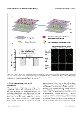
International Journal of Bioprinting                                3D bioprinting for nanoparticle evaluation


            Figure 3. Layer-by-layer 3D skin model for evaluation of skin penetration efficacy of nanoparticles (NPs) with different charges. (A) Three-dimensional
            skin model fabricated through layer-by-layer deposition of collagen and fibroblasts. (B) Amine-modified positively charged polystyrene NPs penetrate
            collagen layers more effectively than hydroxyl and sulfate-modified negatively charged polystyrene NPs. Reproduced with permission from Hou et al.
                                                                                                            56
            Copyright © 2017 Society for Laboratory Automation and Screening.

            4. Three-dimensional bioprinted                    incorporating the patient’s own cartilage cells for better
            ear models                                         integration and functionality post-implantation. 58,59
                                                               Recent studies have successfully integrated microvascular
            Three-dimensional  bioprinting  technology  has    networks within these bioprinted ear models, enhancing
            significantly advanced the development of ear models   tissue viability by mimicking natural nutrient and oxygen
            for tissue engineering and regenerative medicine. This   supply. Additionally, 3D bioprinting has shown potential
            technology allows for the precise deposition of cells and   in incorporating therapeutic agents into the printed
            biomaterials, enabling the creation of complex structures   structures,  improving  treatments  for  conditions  such as
            that closely mimic the natural architecture of human   cartilage damage and ear deformities. These technological
            tissues. The printing of ear constructs, in particular, uses   advancements have revolutionized ear model fabrication,
            patient-specific imaging data to achieve high accuracy,   offering  robust  and  customizable  solutions that enhance


            Volume 10 Issue 5 (2024)                        11                                doi: 10.36922/ijb.4273