
Page 14 - IJB-10-5
P. 14
International Journal of Bioprinting 3D bioprinting for nanoparticle evaluation
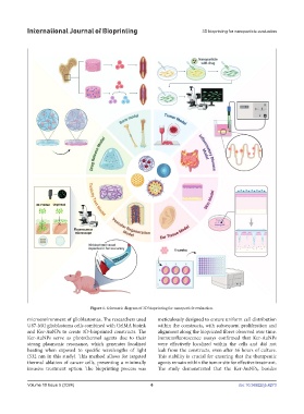
Figure 1. Schematic diagram of 3D bioprinting for nanoparticle evaluation.
microenvironment of glioblastomas. The researchers used meticulously designed to ensure uniform cell distribution
U87-MG glioblastoma cells combined with GelMA bioink within the constructs, with subsequent proliferation and
and Ker-AuNPs to create 3D-bioprinted constructs. The alignment along the bioprinted fibers observed over time.
Ker-AuNPs serve as photothermal agents due to their Immunofluorescence assays confirmed that Ker-AuNPs
strong plasmonic resonance, which generates localized were effectively localized within the cells and did not
heating when exposed to specific wavelengths of light leak from the constructs, even after 56 hours of culture.
(532 nm in this study). This method allows for targeted This stability is crucial for ensuring that the therapeutic
thermal ablation of cancer cells, presenting a minimally agents remain within the tumor site for effective treatment.
invasive treatment option. The bioprinting process was The study demonstrated that the Ker-AuNPs, besides
Volume 10 Issue 5 (2024) 6 doi: 10.36922/ijb.4273