
Page 15 - IJB-10-5
P. 15
International Journal of Bioprinting 3D bioprinting for nanoparticle evaluation
37
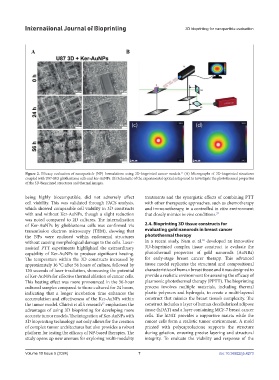
Figure 2. Efficacy evaluation of nanoparticle (NP) formulations using 3D-bioprinted cancer models. (A) Micrographs of 3D-bioprinted structures
coupled with U87-MG glioblastoma cells and ker-AuNPs. (B) Schematic of the experimental optical setup used to investigate the photothermal properties
of the 3D-bioprinted structures and thermal images.
being highly biocompatible, did not adversely affect treatments and the synergistic effects of combining PTT
cell viability. This was validated through FACS analysis, with other therapeutic approaches, such as chemotherapy
which showed comparable cell viability in 3D constructs and immunotherapy, in a controlled in vitro environment
with and without Ker-AuNPs, though a slight reduction that closely mimics in vivo conditions. 37
was noted compared to 2D cultures. The internalization
of Ker-AuNPs by glioblastoma cells was confirmed via 2.4. Bioprinting 3D tissue constructs for
transmission electron microscopy (TEM), showing that evaluating gold nanorods in breast cancer
the NPs were enclosed within endosomal structures photothermal therapy
38
without causing morphological damage to the cells. Laser- In a recent study, Nam et al. developed an innovative
assisted PTT experiments highlighted the extraordinary 3D-bioprinted complex tissue construct to evaluate the
capability of Ker-AuNPs to produce significant heating. photothermal properties of gold nanorods (AuNRs)
The temperature within the 3D constructs increased by for early-stage breast cancer therapy. This advanced
approximately 16 °C after 56 hours of culture, followed by tissue model replicates the structural and compositional
120 seconds of laser irradiation, showcasing the potential characteristics of human breast tissue and it was designed to
of Ker-AuNPs for effective thermal ablation of cancer cells. provide a realistic environment for assessing the efficacy of
This heating effect was more pronounced in the 56-hour plasmonic photothermal therapy (PPTT). The bioprinting
cultured samples compared to those cultured for 24 hours, process involves multiple materials, including thermal
indicating that a longer incubation time enhances the plastic polymers and hydrogels, to create a multi-layered
accumulation and effectiveness of the Ker-AuNPs within construct that mimics the breast tissue’s complexity. The
the tumor model. Chirivì et al.’s research emphasizes the construct includes a layer of human decellularized adipose
37
advantages of using 3D bioprinting for developing more tissue (hDAT) and a layer containing MCF-7 breast cancer
accurate tumor models. The integration of Ker-AuNPs with cells. The hDAT provides a supportive matrix while the
3D bioprinting technology not only allows for the creation cancer cells form a realistic tumor environment. A mold
of complex tumor architectures but also provides a robust printed with polycaprolactone supports the structure
platform for testing the efficacy of NP-based therapies. The during gelation, ensuring precise layering and structural
study opens up new avenues for exploring multi-modality integrity. To evaluate the viability and response of the
Volume 10 Issue 5 (2024) 7 doi: 10.36922/ijb.4273