Page 244 - IJB-10-5
P. 244
International Journal of Bioprinting                                  3D printed hydrogels for tumor therapy


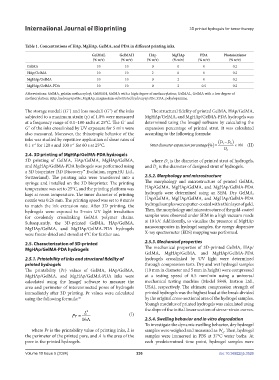
            Table 1. Concentrations of HAp, MgHAp, GelMA, and PDA in different printing inks.
                                        GelMAL      GelMAH       HAp      MgHAp       PDA       Photoinitiator
                                         (% w/v)     (% w/v)    (% w/v)    (% w/v)   (% w/v)       (% w/v)
             GelMA                        10          10          0          0          0           0.2
             HAp/GelMA                    10          10          2          0          0           0.2
             MgHAp/GelMA                  10          10          0          2          0           0.2
             MgHAp/GelMA-PDA              10          10          0          2         0.5          0.2
            Abbreviations: GelMA, gelatin methacryloyl; GelMAH, GelMA with a high degree of methacrylation; GelMAL, GelMA with a low degree of
            methacrylation; HAp, hydroxyapatite; MgHAp, magnesium-substituted hydroxyapatite; PDA, polydopamine.


            The storage moduli (G´) and loss moduli (G˝) of the inks   The structural fidelity of printed GelMA, HAp/GelMA,
            subjected to a maximum strain (γ) of 1.0% were measured   MgHAp/GelMA, and MgHAp/GelMA-PDA hydrogels was
            at a frequency range of 0.1–100 rad/s at 25°C. The G´ and   determined using the ImageJ software by calculating the
            G˝ of the inks crosslinked by UV exposure for 5 min were   expansion  percentage  of  printed  strut.  It  was  calculated
            also measured. Moreover, the thixotropic behavior of the   according to the following formula:
            inks was studied by repetitive application of shear rates of                      ( D − )
                                                                                                  D
            0.1 s for 120 s and 100 s  for 60 s at 25°C.            Strut diameter expansion percentage % () =  1  0  ×100  (II)
                                −1
               −1
                                                                                                D 0
            2.4. 3D printing of MgHAp/GelMA-PDA hydrogels
            3D printing of GelMA, HAp/GelMA, MgHAp/GelMA,         where D  is the diameter of printed strut of hydrogels,
                                                                        1
            and MgHAp/GelMA-PDA hydrogels was performed using   and D  is the diameter of designed strut of hydrogels.
                                                                    0
            a 3D bioprinter (3D Discovery™ Evolution, regenHU Ltd.,
            Switzerland). The printing inks were transferred into a   2.5.2. Morphology and microstructure
            syringe and installed on the 3D bioprinter. The printing   The morphology and microstructure of printed GelMA,
            temperature was set to 25°C, and the printing platform was   HAp/GelMA, MgHAp/GelMA, and MgHAp/GelMA-PDA
            kept at room temperature. The inner diameter of printing   hydrogels were determined using an SEM. Dry GelMA,
            nozzle was 0.26 mm. The printing speed was set to 8 mm/s   HAp/GelMA, MgHAp/GelMA, and MgHAp/GelMA-PDA
            to match the ink extrusion rate. After  3D printing, the   hydrogel samples were sputter-coated with a thin layer of gold.
            hydrogels were exposed to 5-min UV light irradiation   Then, the morphology and microstructure of the gold-coated
            for covalently crosslinking GelMA polymer chains.   samples were observed under SEM in a high vacuum mode
            Subsequently, the 3D-printed GelMA, HAp/GelMA,     at 10 kV. Additionally, to visualize the presence of MgHAp
            MgHAp/GelMA, and MgHAp/GelMA-PDA hydrogels         nanocomposites in hydrogel samples, the energy dispersive
            were freeze-dried and stored at 4°C for further use.   X-ray spectrometer (EDS) mapping was performed.

            2.5. Characterization of 3D-printed                2.5.3. Mechanical properties
            MgHAp/GelMA-PDA hydrogels                          The  mechanical  properties of  3D-printed GelMA,  HAp/
                                                               GelMA, MgHAp/GelMA, and MgHAp/GelMA-PDA
            2.5.1. Printability of inks and structural fidelity of   hydrogels crosslinked by UV light were determined
            printed hydrogels                                  through compression tests. Dry and wet hydrogel samples
            The printability (Pr) values of GelMA, HAp/GelMA,   (10 mm in diameter and 5 mm in height) were compressed
            MgHAp/GelMA, and MgHAp/GelMA-PDA inks were         at a testing speed of 0.5 mm/min using a universal
            calculated using the ImageJ software to measure the   mechanical testing machine (Model 5848, Instron Ltd.,
            area and perimeter of interconnected pores of hydrogels   USA), respectively. The ultimate compression strength of
            immediately after 3D printing. Pr values were calculated   printed hydrogels was the highest load at the break divided
            using the following formula: 38                    by the original cross-sectional area of the hydrogel samples.
                                                               Young’s modulus of printed hydrogels was calculated using
                                                               the slope of the initial linear section of stress–strain curves.
                                      2
                                Pr  =  L                (I)
                                    16A                        2.5.4. Swelling behavior and in vitro degradation
                                                               To investigate the dynamic swelling behavior, dry hydrogel
               where Pr is the printability value of printing inks, L is   samples were weighed and measured as W . Then, hydrogel
                                                                                                0
            the perimeter of the printed pore, and A is the area of the   samples  were  immersed  in  PBS  at  37°C  water  baths.  At
            pore in the printed hydrogels.                     each  predetermined  time  point,  hydrogel samples were

            Volume 10 Issue 5 (2024)                       236                                doi: 10.36922/ijb.3526

