
Page 28 - IJB-10-5
P. 28
International Journal of Bioprinting 3D bioprinting for nanoparticle evaluation
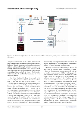
Figure 8. Three-dimensional bioprinted matrix model that closely mimics real tissue environments, providing a more realistic assessment of nanoparticle
(NP) toxicity. 115
compared to conventional 2D cell cultures. The researchers exposed to AgNPs was significantly higher compared to 2D
used a customized 3D bioprinter to print viscous cell-laden cultures, suggesting that the 3D-bioprinted models better
hydrogels. These hydrogels were composed of a mixture of replicate the in vivo responses to NP exposure.
alginate, gelatin, and Matrigel, optimized to support cell The study also emphasizes the advantages of using 3D
viability and structural integrity. The printed scaffolds bioprinting for nanotoxicology investigations. Firstly,
113
115
were then used to culture immortalized lung cell lines over it allows for the creation of more physiologically relevant
extended periods, specifically to observe the interaction models that can maintain cell viability over longer periods
with NPs such as 40 nm latex-fluorescent and 11–14 nm without frequent passages. Secondly, the ability of NPs to
silver NPs (AgNPs). diffuse and interact with cells in a 3D matrix closely mimics
One of the significant findings of this study is the real tissue environments. Thirdly, the 3D-printed scaffolds
enhanced cell proliferation within the 3D-printed scaffolds. provide a more realistic assessment of NP toxicity, showing
5
Over 14 days, the cell count increased from 5 × 10 to similar toxicological responses to those observed in in vivo
6
1.27 × 10 , demonstrating a conducive environment for cell studies, such as those with zebrafish, insects, and rodents.
114
growth. Furthermore, the lipid peroxidation levels were In summary, the research conducted by Gerbolés et al.
115
reduced by 91%, indicating a decrease in oxidative stress, demonstrates that 3D bioprinting of organoid-based
which is a common response to NP exposure. The 3D scaffolds presents a promising advancement for the long-
environment also showed minimal cell death over a 21-day term investigation of NP toxicology. This method not only
period, highlighting the protective nature of the bioprinted improves the accuracy of in vitro models but also reduces the
scaffolds. The diffusion of NPs within the 3D scaffolds was use of animals in experiments, aligning with the principles
another critical observation. Fluorescent NPs were able to of the 3Rs (Replacement, Reduction, and Refinement)
spread throughout the scaffolds, unlike in the unprinted in toxicological research. The findings suggest that 3D
environments. This characteristic is vital for mimicking bioprinting could be a vital tool for future nanotoxicology
in vivo conditions where NPs interact with cells in a more and nanomedicine research, providing more reliable and
complex 3D space. Additionally, cell viability in 3D cultures ethically sound alternatives to traditional methods. 115
Volume 10 Issue 5 (2024) 20 doi: 10.36922/ijb.4273