
Page 311 - IJB-10-5
P. 311
International Journal of Bioprinting Proteins-loaded 3D-printed PEEK cage
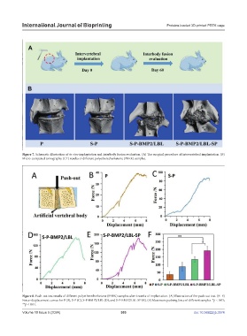
Figure 7. Schematic illustration of in vivo implantation and interbody fusion evaluation. (A) The surgical procedure of intervertebral implantation. (B)
Micro-computed tomography (CT) results of different polyetheretherketone (PEEK) samples.
Figure 8. Push-out test results of different polyetheretherketone (PEEK) samples after 6 weeks of implantation. (A) Illustration of the push-out test. (B–E)
Force–displacement curves for P (B), S-P (C), S-P-BMP2/LBL (D), and S-P-BMP2/LBL-SP (E). (F) Maximum pushing force of different samples. *p < 0.05;
**p < 0.01.
Volume 10 Issue 5 (2024) 303 doi: 10.36922/ijb.3574