
Page 309 - IJB-10-5
P. 309
International Journal of Bioprinting Proteins-loaded 3D-printed PEEK cage
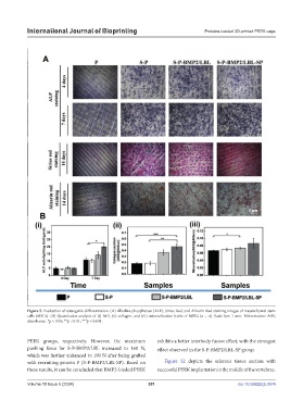
Figure 5. Evaluation of osteogenic differentiation. (A) Alkaline phosphatase (ALP), Sirius Red, and Alizarin Red staining images of mesenchymal stem
cells (MSCs). (B) Quantitative analysis of (i) ALP, (ii) collagen, and (iii) mineralization levels of MSCs (n = 4). Scale bar: 1 mm. Abbreviation: ABS,
absorbance. *p < 0.05; **p < 0.01; ***p < 0.001.
PEEK groups, respectively. However, the maximum exhibits a better interbody fusion effect, with the strongest
pushing force for S-P-BMP2/LBL increased to 160 N, effect observed in the S-P-BMP2/LBL-SP group.
which was further enhanced to 190 N after being grafted
with recruiting protein P (S-P-BMP2/LBL-SP). Based on Figure S2 depicts the sclerous tissue section with
these results, it can be concluded that BMP2-loaded PEEK successful PEEK implantation in the middle of the vertebrae.
Volume 10 Issue 5 (2024) 301 doi: 10.36922/ijb.3574