
Page 305 - IJB-10-5
P. 305
International Journal of Bioprinting Proteins-loaded 3D-printed PEEK cage
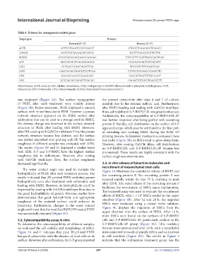
Table 1. Primers for osteogenesis-related genes
Target gene Primers
Forward (5’–3’) Reverse (5’–3’)
ACTB ATTTCTGAATGGCCCAGGT CTGCCTCAACACCTCAACC
GAPDH GCTCTCCAGAACATCATCC TGCTTCACCACCTTCTTG
RUNX2 GCCGTAGAGAGCAGGGAAGAC CTGGCTTGGATTAGGGAGTCAC
ALP AGCGACACGGACAAGAAGC GGCAAAGACCGCCACATC
COLI CCTGAGCCAGCAGATTGA TCCGCTCTTCCAGTCAG
OCN GAGGGCAGTAAGGTGGTGAA COTCCTGGAAGCCAATGTG
OPN GACAGCAACGGGAAGACC CAGGCTGGCTTTGGAACT
OPG GCCCAGACGAGATTGAGAG CAGACTGTGGGTGACGGTT
Abbreviations: ACTB, Actin-β; ALP, Alkaline phosphatase; COLI, Collagen type I; GAPDH, Glyceraldehyde 3-phosphate dehydrogenase; OCN,
Osteocalcin; OPN, Osteopontin; OPG, Osteoprotegerin; RUNX2, Runt-related transcription factor 2.
was employed (Figure 2A). The surface topographies the poorest cytoactivity after days 4 and 7 of culture,
of PEEK after each treatment were notably altered possibly due to the residual sulfuric acid. Furthermore,
(Figure 2B). Before treatment, PEEK displayed a smooth after BMP2-loading and sealing with Gel/Chi multilayer
surface, with minor lines due to FDM. However, a porous films, cell viability in S-P-BMP2/LBL was greatly enhanced.
network structure appeared on the PEEK surface after Additionally, the cytocompatibility of S-P-BMP2/LBL-SP
sulfonation that can be used as a storage pool for BMP2. was further improved after being grafted with recruiting
No obvious change was observed in the surface network protein P. Notably, cell distribution on the surface of S-P
structure of PEEK after loading with BMP2. However, appeared stripy, which may be attributed to the line path
after LBL coating with Gel/Chi multilayer films, the porous of extruding and stacking PEEK during the FDM 3D
network structure became less distinct, and the surface printing process. Sulfonation treatment accentuated these
was further smoothed after grafting with SP. The surface line paths (Figure 3B), as MSCs tend to grow along them.
roughness of different samples was evaluated with AFM. However, after coating Gel/Chi films, cell distributions
The results (Figure 2D and E) displayed a similar trend on S-P-BMP2/LBL and S-P-BMP2/LBL-SP became less
with SEM. S-P and S-P-BMP2 have the highest surface pronounced. These results are highly consistent with the
roughness due to sulfonation. However, after coating surface roughness observations.
with Gel/Chi multilayer films, the surface roughness
decreased significantly. 3.3. In vitro release of bioactive molecules and
recruitment of mesenchymal stem cells
The water contact angle was used to evaluate the Figure 4A illustrates the cumulative release of BMP2 and
hydrophilicity of PEEK after each treatment process. The the recruiting protein P. The recruiting protein P was
results indicated that 3D-printed PEEK exhibited poorer released rapidly within the first 72 h, reaching its peak
hydrophilicity even after treatment with sulfonation and after 120 h. The rapid release of the recruiting protein P
loading with BMP2. However, its hydrophilicity could be facilitates the recruitment of MSCs upon implantation.
improved by coating with Gel/Chi multilayer films due to The transwell assay was used to evaluate the recruitment
the good hydrophilicity of gelatin. Previous studies have effects of BMP2, with 1 × 10 MSCs seeded in the upper
4
demonstrated that good hydrophilicity and appropriate chamber (Figure 4B). After 12 and 24 h, the migrated
roughness of the material surface could enhance its
bioactivity. Furthermore, changes in the water contact MSCs were evaluated using a crystal violet solution.
angle confirmed that the modified BMP2/SP-loaded PEEK Figure 4C displays the migration of MSCs from the
was successfully prepared (Figure 2C). upper chamber onto the surface of PEEK. After 12 h,
more MSCs were found on the surfaces of S-P-BMP2/
3.2. Cytocompatibility assay in vitro LBL and S-P-BMP2/LBL-SP, particularly evident in the
To determine the cytocompatibility of different samples, S-P-BMP2/LBL-SP group (Figure 4C). This tendency
we evaluated the cell viability and morphology of MSCs. became more pronounced after 24 h, with a remarkable
Figure 3A and C indicates that pure 3D-printed PEEK interconnected network of purple MSCs and an increase
has good cytoactivity, with the absence of dead cells on its in cell numbers (Figure 4D). The results in Figure 3
surface. However, after sulfonation, the S-P group reported indicate that the sulfonation treatment group has the
Volume 10 Issue 5 (2024) 297 doi: 10.36922/ijb.3574