
Page 303 - IJB-10-5
P. 303
International Journal of Bioprinting Proteins-loaded 3D-printed PEEK cage
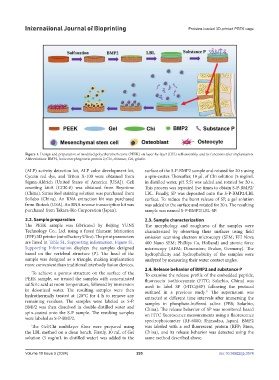
Figure 1. Design and preparation of modified polyetheretherketone (PEEK) via layer-by-layer (LBL) self-assembly, and its functions after implantation.
Abbreviations: BMP2, bone morphogenetic protein 2; Chi, chitosan; Gel, gelatin.
(ALP) activity detection kit, ALP color development kit, surface of the S-P-BMP2 sample and rotated for 20 s using
Cycein red dye, and Triton X-100 were obtained from a spin-coater. Thereafter, 10 μL of Chi solution (5 mg/mL
Sigma-Aldrich (United States of America [USA]). Cell in distilled water, pH 5.5) was added and rotated for 20 s.
counting kit-8 (CCK-8) was obtained from Beyotime This process was repeated five times to obtain S-P-BMP2/
(China). Sirius Red staining solution was purchased from LBL. Finally, SP was deposited onto the S-P-BMP2/LBL
Sollebo (China). An RNA extraction kit was purchased surface. To reduce the burst release of SP, a gel solution
from Bioteck (USA). An RNA reverse transcription kit was was added to the surface and rotated for 20 s. The resulting
purchased from Takara-Bio Corporation (Japan). sample was named S-P-BMP2/LBL-SP.
2.2. Sample preparation 2.3. Sample characterization
The PEEK sample was fabricated by Beijing YUNS The morphology and roughness of the samples were
Technology Co., Ltd. using a fused filament fabrication characterized by observing their surfaces using field
(FFF) 3D printer (miniFactory Ultra). The print parameters emission scanning electron microscopy (SEM; FEI Nova
are listed in Table S1, Supporting information. Figure S1, 400 Nano SEM; Phillips Co, Holland) and atomic force
Supporting Information displays the samples designed microscopy (AFM; Dimension; Bruker, Germany). The
based on the vertebral structure (P). The head of the hydrophilicity and hydrophobicity of the samples were
sample was designed as a triangle, making implantation analyzed by measuring their water contact angles.
more convenient than traditional interbody fusion devices.
2.4. Release behavior of BMP2 and substance P
To achieve a porous structure on the surface of the To examine the release profile of the embedded peptide,
PEEK sample, we treated the samples with concentrated fluorescein isothiocyanate (FITC; Solarbio, China) was
sulfuric acid at room temperature, followed by immersion used to label SP (FITC@SP) following the protocol
in deionized water. The resulting samples were then outlined in a previous study. The supernatant was
31
hydrothermally treated at 120°C for 4 h to remove any extracted at different time intervals after immersing the
remaining residues. The samples were labeled as S-P. samples in phosphate-buffered saline (PBS; Solarbio,
BMP2 was then dissolved in double-distilled water and China). The release behavior of SP was monitored based
spin-coated onto the S-P sample. The resulting samples on FITC fluorescence measurements using a fluorescence
were labeled as S-P-BMP2. spectrophotometer (RF-6000; Shimadzu, Japan). BMP2
The Gel/Chi multilayer films were prepared using was labeled with a red fluorescent protein (RFP; Bioss,
the LBL method on a clean bench. Firstly, 10 mL of Gel China), and its release behavior was detected using the
solution (5 mg/mL in distilled water) was added to the same method described above.
Volume 10 Issue 5 (2024) 295 doi: 10.36922/ijb.3574