
Page 334 - IJB-10-5
P. 334
International Journal of Bioprinting Immunomodulatory bone repair by MBG/PCL
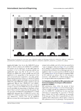
Figure 2. Surface morphology and contact angle images of MBG/PCL scaffolds. (A) SEM images of 0MBG/PCL, 5MBG/PCL, 10MBG/PCL, 20MBG/PCL,
and 30MBG/PCL scaffolds. (B) Contact angle pictures of 0MBG/PCL, 5MBG/PCL, 10MBG/PCL, 20MBG/PCL, and 30MBG/PCL scaffolds.
significantly higher than that in the 0MBG/PCL group, cultured with scaffolds, and ALP activity was quantified at
while the difference between the 30MBG/PCL and 0MBG/ 7 days of osteogenic induction (Figure 4D), and scaffolds
PCL groups was not significant. On the other hand, the in the 10MBG/PCL group had the highest ALP expression,
expression of Col1 in the MBG-containing scaffolds was while the difference between the remaining groups was not
significantly higher than that in the pure PCL group. significant, which was also in agreement with the results of
Notably, the 10MBG/PCL group scaffolds were the most ALP staining (Figure 4B). In summary, the doping of MBG
significant in the upregulation of the expression of the in PCL materials can enhance their in vitro osteogenic
above-mentioned genes compared to the other groups. properties, with 10MBG/PCL scaffolds having the best
Regarding the expression of Runx2, the 10MBG/PCL group pro-osteogenic differentiation properties for BMSCs.
likewise exhibited significantly higher expression than the
0MBG/PCL and 5MBG/PCL groups, whereas it was not 3.4. Evaluation of scaffolds for MPs polarization and
significantly different than 20MBG/PCL and 30MBG/ immunomodulation of osteogenic differentiation
PCL in terms of Runx2 expression. Even after 14 days of properties of BMSCs
osteogenic induction, the 10MBG/PCL group still held the MPs polarization status affects the process of bone
most potent ability in upregulating the expression of Opn, regeneration. Based on Figure 5A, we found that among the
Runx2, Bmp2, and Col1 compared to the other groups. The various scaffolds inducing the polarization of RAW264.7
Alp expression of 10MBG/PCL and 20MBG/PCL scaffolds cells, the 10MBG/PCL scaffold had the most significant
significantly surpassed that of 0MBG/PCL group, with performance in upregulating the expression of M2-type
10MBG/PCL exhibiting higher gene expression compared genes (CD206, Arg) as well as inhibiting the expression
with 30MBG/PCL, whereas there was no significant of M1-type genes (Tnfa, Il1b). Compared with pure PCL
difference between the remaining groups. BMSCs were co- scaffolds, MBG endowed PCL with an ability to promote
MPs polarization to the M2 phenotype. The expression of
Volume 10 Issue 5 (2024) 326 doi: 10.36922/ijb.3551