
Page 479 - IJB-10-5
P. 479
International Journal of Bioprinting 3D-printed post-otoplasty ear retainer
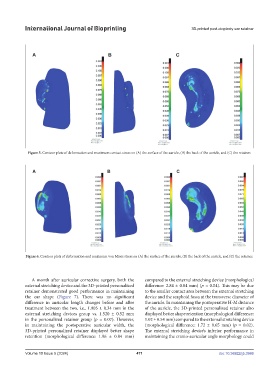
Figure 5. Contour plots of deformation and maximum contact stress on (A) the surface of the auricle, (B) the back of the auricle, and (C) the retainer.
Figure 6. Contour plots of deformation and maximum von Mises stress on (A) the surface of the auricle, (B) the back of the auricle, and (C) the retainer.
A month after auricular corrective surgery, both the compared to the external stretching device (morphological
external stretching device and the 3D-printed personalized difference: 2.84 ± 0.84 mm) (p = 0.04). This may be due
retainer demonstrated good performance in maintaining to the smaller contact area between the external stretching
the ear shape (Figure 7). There was no significant device and the scaphoid fossa at the transverse diameter of
difference in auricular length changes before and after the auricle. In maintaining the postoperative H-M distance
treatment between the two, i.e., 1.905 ± 0.34 mm in the of the auricle, the 3D-printed personalized retainer also
external stretching devices group vs. 1.520 ± 0.52 mm displayed better shape retention (morphological difference:
in the personalized retainer group (p = 0.07). However, 1.02 ± 0.54 mm) compared to the external stretching device
in maintaining the postoperative auricular width, the (morphological difference: 1.72 ± 0.65 mm) (p = 0.02).
3D-printed personalized retainer displayed better shape The external stretching device’s inferior performance in
retention (morphological difference: 1.96 ± 0.84 mm) maintaining the cranio-auricular angle morphology could
Volume 10 Issue 5 (2024) 471 doi: 10.36922/ijb.3986