
Page 53 - IJB-6-1
P. 53
Gantumur, et al.
A lysis. The possible reason for cell death might be
the mechanical stress from the high concentration
of CNF incorporated in the hydrogels during the
mixing procedure and the culture time. In the
previous study that also used CNF for the extrusion
system, low cell viability (<70%) for human
B chondrocytes, which has different morphology
than our model cell line, was reported. It could be
considered as one of the drawbacks of using CNF
for bioink . Although it is out of the scope of this
[35]
paper, CNF-based bioinks may be more suitable
for the regeneration of cartilage tissues than the
other tissues [40,42] . Besides this effect of CNF on
cells, the proposed hydrogelation method can be
C
utilized for 3D bioprinting of living cells.
3.3 Switchable construct surface
3D-printed hydrogel constructs can also be used
as scaffolds for cell culture [43,44] . Since it is well-
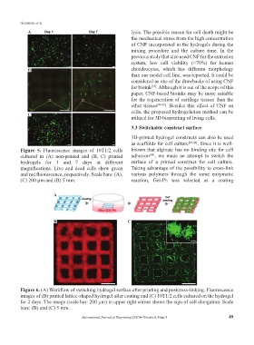
Figure 5. Fluorescence images of 10T1/2 cells known that alginate has no binding site for cell
cultured in (A) non-printed and (B, C) printed adhesion , we made an attempt to switch the
[45]
hydrogels for 1 and 7 days at different surface of a printed construct for cell culture.
magnifications. Live and dead cells show green Taking advantage of the possibility to cross-link
and red fluorescence, respectively. Scale bars: (A), various polymers through the same enzymatic
(C) 200 µm and (B) 5 mm. reaction, Gel-Ph was selected as a coating
A
B C
Figure 6. (A) Workflow of switching hydrogel surface after printing and postcross-linking. Fluorescence
images of (B) printed lattice-shaped hydrogel after coating and (C) 10T1/2 cells cultured on the hydrogel
for 2 days. The image (scale bar: 200 µm) in upper right corner shows the sign of cell elongation. Scale
bars: (B) and (C) 5 mm.
International Journal of Bioprinting (2020)–Volume 6, Issue 1 49