
Page 54 - IJB-6-1
P. 54
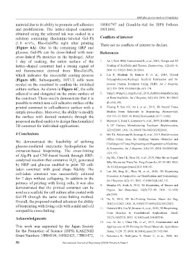
HRP plus glucose-mediated bioprinting
           material due to its ability to promote cell adhesion   18H01797 and Grand-In-Aid for  JSPS Fellows
           and proliferation.  The lattice-shaped construct    18J11601.
           obtained using the selected ink was soaked in a
           solution containing rhodamine-labeled Gel-Ph        Conflicts of interest
           (1.0 w/v%, Rho-Gel-Ph) right after printing         There are no conflicts of interest to declare.
           (Figure 6A). Due to the remaining HRP and
           glucose, Gel-Ph can be cross-linked with non-       References
           cross-linked Ph moieties in the hydrogel. After
           1  day of  soaking, the entire  surface of  the     1.   An J, Teoh JEM, Suntornnond R, et al., 2015, Design and 3D
           lattice-shaped construct had a strong signal of         Printing of Scaffolds and Tissues. Engineering, 1(2):261–8.
           red  fluorescence  derived  from  Rho-Gel-Ph,           DOI 10.15302/J-ENG-2015061.
           which indicates the successful coating process      2.   Liu F, Mishbak H, Bartolo P,  et al., 2019, Hybrid
           (Figure  6B).  Subsequently,  10T1/2  cells  were       Polycaprolactone/Hydrogel  Scaffold  Fabrication  and  In-
           seeded on the construct to confirm the switched         process Plasma  Treatment  Using PABS.  Int J Bioprint,
           culture surface. As shown in Figure 6C, the cells       5(1):174. DOI: 10.18063/ijb.v5i1.174.
           adhered to and elongated on the entire surface of   3.   Yang Y, Wang G, Liang H, et al., 2019, Additive Manufacturing
           the construct. These results demonstrate that it is     of Bone Scaffolds. Int J Bioprint, 5(1):148. DOI: 10.18063/
           possible to switch non-cell-adhesive surface of the     ijb.v5i1.148.
           printed construct to cell-adhesive surface with a   4.   Zhuang  P,  Sun AX, An  J,  et al., 2018, 3D  Neural  Tissue
           simple procedure. Moreover, the ability to modify       Models: From Spheroids to Bioprinting.  Biomaterials,
           the surface with desired materials through the          154:113–33. DOI: 10.1016/j.biomaterials.2017.10.002.
           proposed method enables to design functionalized    5.   Mironov V, Trusk T, Kasyanov V, et al., 2009, Biofabrication:
           3D construct for individual applications.               A  21   Century Manufacturing Paradigm.  Biofabrication,
                                                                       st
                                                                   1(2):022001. DOI: 10.1088/1758-5082/1/2/022001.
           4 Conclusions                                       6.   Mir TA, Nakamura M, Iwanaga S, et al., 2019, Biofabrication
                                                                   Offers Future Hope for  Tackling  Various Obstacles and
           We demonstrated  the feasibility  of utilizing          Challenges in Tissue Engineering and Regenerative Medicine:
           glucose-mediated  enzymatic  hydrogelation  for         A Perspective, Int J Bioprint, 5(1):153. DOI: 10.18063/ijb.
           extrusion-based  bioprinting.  The  cross-linking       v5i1.153.
           of Alg-Ph and CNF-based bioink through HRP-         7.   Ng WL, Chua CK, Shen YF, et al., 2019, Print Me an Organ!
           catalyzed reaction that consumes H O  generated         Why We are not There Yet. Prog Polym Sci, 97:101145. DOI:
                                             2
                                                2
           by HRP and glucose enabled to print 3D cell-            10.1016/j.prog-polymsci.2019.101145.
           laden  construct  with  good  shape  fidelity.  The   8.   Lee  JM,  Sing  SL,  Zhou  M,  et al., 2018, 3D Bioprinting
           cell-laden  construct was successfully cultured         Processes: A Perspective on Classification and Terminology.
           for 7 days without collapsing. In addition to the       Int J Bioprint, 4(2):151. DOI: 10.18063/ijb.v4i2.151.
           potency of printing with living cells, it was also
           demonstrated  that  the printed construct  can be   9.   Murphy SV, Atala A, 2014, 3D Bioprinting of Tissues and
           used as a scaffold for cell culture after coated with   Organs.  Nat Biotechnol, 32(8):773–85. DOI:  10.1038/
                                                                   nbt.2958.
           Gel-Ph through the same cross-linking method.       10.  Du X, 2018, 3D Bio-Printing Review.  Mater Sci Eng,
           Overall, the proposed method advances the ability       301(1):012023. DOI: 10.1088/1757-899X/301/1/012023.
           of bioprinting with living cells with a mild and cell   11.  Heinrich MA, Liu W, Jimenez A, et al., 2019, 3D Bioprinting:
           compatible cross-linking.
                                                                   From  Benches  to  Translational  Applications.  Small,
           Acknowledgments                                         15(23):1805510. DOI: 10.1002/smll.201805510.
                                                               12.  Lee JY,  An J, Chua CK,  et al., 2017, Fundamentals  and
           This work was supported by the Japan Society            Applications of 3D Printing for Novel Materials. Appl Mater
           for the Promotion of Science (JSPS) KAKENHI             Today, 7:120–33. DOI: 10.1016/j.apmt.2017.02.004.
           Grant Numbers 15H04194, 16H02423, 17H03472,         13.  Nakamura  K, Nishiyama  Y, Henmi C,  et al., 2008, Ink

           50                          International Journal of Bioprinting (2020)–Volume 6, Issue 1