
Page 59 - IJB-6-1
P. 59
Huyan, et al.
hydrogel extract constituted the control. 10 μl cell density of 1 × 10 cells/ml. The combination
6
CCK-8 solutions were added to each well of the of cells and hydrogel was used to print the 3D
plate and incubated at 37°C for 4 h. Absorbance structure, which was then cultured in a CO
2
at a wavelength of 450 nm was measured using incubator at 5% CO , 37°C , and evaluated using
[18]
2
a microplate reader. All results are presented as live and dead staining of the cells on days 1, 4,
optical density (OD) values minus the absorbance and 7. Cell growth was observed using a Laser
of blank wells. The distribution of cells was Confocal Microscope (Nikon A1). Live cells
observed using fluorescence microscopy. appeared green and dead cells red.
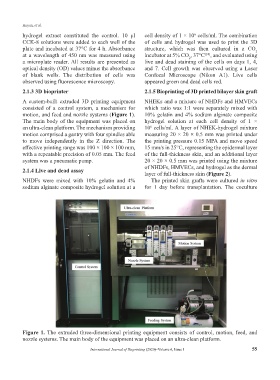
2.1.3 3D bioprinter 2.1.5 Bioprinting of 3D printed bilayer skin graft
A custom-built extruded 3D printing equipment NHEKs and a mixture of NHDFs and HMVECs
consisted of a control system, a mechanism for which ratio was 1:1 were separately mixed with
motion, and feed and nozzle systems (Figure 1). 10% gelatin and 4% sodium alginate composite
The main body of the equipment was placed on hydrogel solution at each cell density of 1 ×
an ultra-clean platform. The mechanism providing 10 cells/ml. A layer of NHEK-hydrogel mixture
6
motion comprised a gantry with four spindles able measuring 20 × 20 × 0.5 mm was printed under
to move independently in the Z direction. The the printing pressure 0.15 MPA and move speed
effective printing range was 100 × 100 × 100 mm, 15 mm/s in 25°C, representing the epidermal layer
with a repeatable precision of 0.05 mm. The feed of the full-thickness skin, and an additional layer
system was a pneumatic pump. 20 × 20 × 0.5 mm was printed using the mixture
of NHDFs, HMVECs, and hydrogel as the dermal
2.1.4 Live and dead assay
layer of full-thickness skin (Figure 2).
NHDFs were mixed with 10% gelatin and 4% The printed skin grafts were cultured in vitro
sodium alginate composite hydrogel solution at a for 1 day before transplantation. The coculture
Figure 1. The extruded three-dimensional printing equipment consists of control, motion, feed, and
nozzle systems. The main body of the equipment was placed on an ultra-clean platform.
International Journal of Bioprinting (2020)–Volume 6, Issue 1 55