
Page 64 - IJB-6-1
P. 64
Vascularization of printed bilayer skin grafts
A B
C D
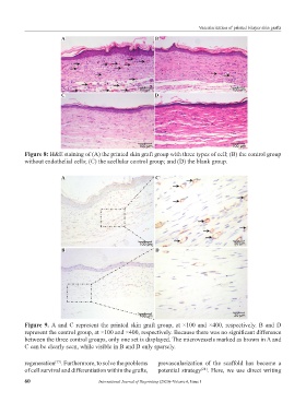
Figure 8: H&E staining of (A) the printed skin graft group with three types of cell; (B) the control group
without endothelial cells; (C) the acellular control group; and (D) the blank group.
A C
B D
Figure 9. A and C represent the printed skin graft group, at ×100 and ×400, respectively. B and D
represent the control group, at ×100 and ×400, respectively. Because there was no significant difference
between the three control groups, only one set is displayed. The microvessels marked as brown in A and
C can be clearly seen, while visible in B and D only sparsely.
regeneration . Furthermore, to solve the problems prevascularization of the scaffold has become a
[27]
of cell survival and differentiation within the grafts, potential strategy . Here, we use direct writing
[28]
60 International Journal of Bioprinting (2020)–Volume 6, Issue 1