
Page 65 - IJB-6-1
P. 65
Huyan, et al.
A C
B D
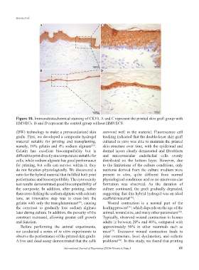
Figure 10. Immunohistochemical staining of CK10. A and C represent the printed skin graft group with
HMVECs. B and D represent the control group without HMVECS.
(DW) technology to make a prevascularized skin survived well in the material. Fluorescence cell
grafts. First, we developed a composite hydrogel tracking indicated that the double-layer skin graft
material suitable for printing and transplanting, cultured in vitro was able to maintain the printed
namely, 10% gelatin and 4% sodium alginate . skin structure over time, with the epidermal and
[29]
Gelatin has excellent biocompatibility but is dermal layers clearly demarcated and fibroblasts
difficult to print directly at a temperature suitable for and microvascular endothelial cells evenly
cells, while sodium alginate has good performance distributed on the bottom layer. However, due
for printing, but cells can survive within it, they to the limitations of the culture conditions, only
do not function physiologically. We discovered a nutrients derived from the culture medium were
ratio for the hybrid material that fulfilled both print present in vitro, quite different from normal
performance and biocompatibility. The cytotoxicity physiological conditions and so no microvascular
test results demonstrated good biocompatibility of formation was observed. As the duration of
the composite. In addition, after printing, rather culture continued, the graft gradually degraded,
than cross-linking the sodium alginate with calcium suggesting that this hybrid hydrogel was an ideal
ions, an innovative step was to cross-link the scaffold material .
[30]
gelatin with only the transglutaminase , causing Wound contraction is a normal part of the
[29]
the construct to gradually lose sodium alginate healing process , which depends on the age of the
[31]
later during culture. In addition, the porosity of the animal, wound size, and many other parameters .
[32]
construct increased, allowing greater cell growth Typically, observed wound contraction in human
and function. adults is between 20% and 40%, compared with
Before performing the animal experiments, approximately 90% in other mammals such as
we conducted a series of in vitro experiments to mice . Excessive wound contraction leads to
[33]
observe the performance of the printed skin grafts. joint contracture, local dysfunction, and esthetic
A live and dead assay demonstrated that the cells problems . In this study, we found that printing
[34]
International Journal of Bioprinting (2020)–Volume 6, Issue 1 61