
Page 60 - IJB-6-1
P. 60
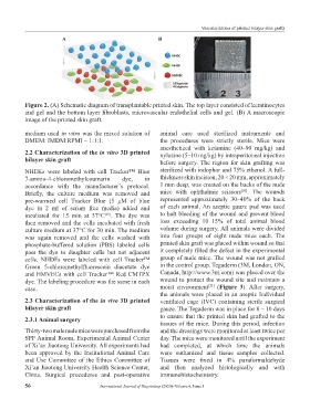
Vascularization of printed bilayer skin grafts
                         A                                    B


           Figure 2. (A) Schematic diagram of transplantable printed skin. The top layer consisted of keratinocytes
           and gel and the bottom layer fibroblasts, microvascular endothelial cells and gel. (B) A macroscopic
           image of the printed skin graft.

           medium used in vitro was the mixed solution of      animal  care  used sterilized  instruments  and
           DMEM: IMDM:RPMI = 1:1:1.                            the procedures were strictly sterile. Mice were
                                                               anesthetized  with ketamine (40–90  mg/kg) and
           2.2 Characterization of the in vitro 3D printed     xylazine (5–10 mg/kg) by intraperitoneal injection
           bilayer skin graft                                  before surgery. The region for skin grafting was

           NHEKs were labeled  with cell  Tracker™  Blue       sterilized with iodophor and 75% ethanol. A full-
           7-amino-4-chloromethylcoumarin       dye,     in    thickness skin incision, 20 × 20 mm, approximately
           accordance  with  the  manufacturer’s protocol.     1 mm deep, was created on the backs of the nude
                                                                                              [20]
           Briefly,  the  culture  medium  was  removed  and   mice with ophthalmic  scissors . The  wounds
           pre-warmed  cell  Tracker  Blue  (5  μM  of  blue   represented  approximately 30–40%  of the  back
           dye in 2  ml of serum free media)  added and        of each animal. An aseptic gauze pad was used
           incubated  for 15  min  at  37°C .  The  dye  was   to halt bleeding of the wound and prevent blood
                                         [19]
           then removed and the cells incubated with fresh     loss exceeding 10–15% of total animal  blood
           culture medium at 37°C for 30 min. The medium       volume during surgery. All animals were divided
           was again removed and the cells washed with         into  four groups of eight  nude  mice  each.  The
           phosphate-buffered solution (PBS) labeled  cells    printed skin graft was placed within wound so that
           pass the dye to daughter cells  but not adjacent    it completely filled the defect in the experimental
           cells.  NHDFs  were  labeled  with  cell  Tracker™   group of nude mice. The wound was not grafted
           Green  5-chloromethylfluorescein  diacetate  dye    in the control group. Tegaderm (3M, London, ON,
           and HMVECs with cell Tracker™ Red CMTPX             Canada, http://www.3m.com) was placed over the
           dye. The labeling procedure was the same in each    wound to protect the wound site and maintain a
                                                                                 [21]
           case.                                               moist  environment  (Figure  3). After  surgery,
                                                               the animals were placed in an aseptic Individual
           2.3 Characterization of the in vivo 3D printed      ventilated cage (IVC) containing sterile surgical
           bilayer skin graft                                  gauze. The Tegaderm was in place for 8 – 10 days
                                                               to ensure that the printed skin had grafted to the
           2.3.1 Animal surgery
                                                               tissues of the mice. During this period, infection
           Thirty-two male nude mice were purchased from the   and the dressings were monitored at least twice per
           SPF Animal Room, Experimental Animal Center         day. The mice were monitored until the experiment
           of Xi’an Jiaotong University. All experiments had   had  completed,  at  which time  the  animals
           been approved by the Institutional Animal Care      were euthanized  and tissue samples collected.
           and Use Committee of the Ethics Committee of        Tissues  were  fixed  in  4%  paraformaldehyde
           Xi’an Jiaotong University Health Science Center,    and  then  analyzed  histologically  and  with
           China. Surgical  procedures and post-operative      immunohistochemistry.

           56                          International Journal of Bioprinting (2020)–Volume 6, Issue 1