
Page 61 - IJB-6-1
P. 61
Huyan, et al.
A B C D
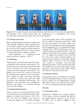
Figure 3. The surgical procedure involved establishing a full-thickness skin wound and transplantation.
(A) Marking of the wound incision lines; (B) establishment of a full-thickness skin wound;
(C) placement of the printed skin graft; (D) covering by Tegaderm.
2.3.2 Wound contraction of 10 mM sodium citrate, 0.05% polysorbate 20,
pH 6.0, cooled for 30 min and washed with 1 × PBS
Mice were photographed on the day of surgery and to recover the antigens. The tissue samples were
at the end of the experiment. A ruler was placed next placed in 100 μl normal goat serum and incubated
to the wound to ensure wounds matched the graft at room temperature for 1 h. An appropriate primary
size. The area of the wound at each time point was antibody (CD31 YM6277 Immunoway, and CK10
measured using ImageJ software . The percentage YM6622 Immunoway) diluted in PBS (1: 500) was
[22]
of wound contraction was defined as follows: added to each sample and incubated overnight at
wound contraction = (1 − wound area at end 4°C. The samples were washed 5 times with PBS
point/wound area at surgery) × 100% . for 5 min each to remove unbound antibody. The
[23]
2.3.3 Histology samples were incubated with a secondary antibody
diluted in PBS (1: 500) for 30 min. The samples
Tissue samples were harvested at specified times, were washed in accordance with previously
including the regenerated and contracted skin. The published procedures then incubated with DAB.
[25]
tissue samples were embedded in paraffin wax and The samples were mounted with neutral chewing
cut into 6 μm-thick slices with a microtome then gum and covered with a coverslip. Finally, staining
stained with hematoxylin and eosin (H&E) . The was observed using a light microscope.
[24]
samples were sealed with a glass coverslip and
the growth and scar formation of the regenerated 2.4 Statistical analysis
skin evaluated by light microscopy, including All quantitative data were calculated as arithmetic
the formation of microvessels and other skin means and standard deviations. A student’s t-test was
accessories, and epidermal differentiation. The used to compare the skin transplantation samples
thickness of the regenerated skin, including the and control groups at the different time periods.
epidermis and dermis, was measured through P < 0.05 was considered statistically significant.
cross-sectional staining.
3 Results
2.3.4 Immunohistochemistry
3.1 Cytotoxicity assay
Tissue sections for immunohistochemistry (IHC)
were dewaxed in xylene and hydrated in a decreasing Cytotoxicity evaluation was conducted using a
gradient of ethanol concentrations (100%, 95%, CCK-8 assay to directly determine the effect of
70%, 50%, and 0%). Samples were boiled for the gelatin-alginate composite on the activity
15 min in an antigen retrieval solution consisting of NHDF cells. As shown in Figure 4, there
International Journal of Bioprinting (2020)–Volume 6, Issue 1 57