
Page 63 - IJB-6-1
P. 63
Huyan, et al.
transplantation group and the control group significant angiogenesis but also that the
without endothelial cells. microvascular-derived cells that were derived
from the printed microvascular endothelial cells,
3.5 Histology confirming that the printed skin graft was capable
H&E staining of the different groups 4 weeks of growing efficiently and promoting its integration
after surgery is displayed in Figure 8. Histological with the mouse tissues and regeneration of blood
analysis demonstrated that the printed skin graft vessels. Figure 10 displays immunohistochemical
group correctly contained all the graft components, staining of Cytokeratin 10(CK10) 4 weeks after
with epidermal and dermal structures that were surgery. CK10 identifies the spinous layer of
intact. Growth of the printed skin graft group the epidermis and represents one of its major
was significantly better than those of the control components. Epidermal growth of the printed skin
groups. First, there was significant angiogenesis grafting group was significantly greater than those
in the dermis, while there were barely any of the control group, with a spinous layer that was
microvessels in the other groups. Second, the significantly thickened.
epidermal layer thickness of this group was also 4 Discussion
significantly greater than the others, a difference
that was significant. The principal purpose of this study was to evaluate
3.6 Immunohistochemistry the capability of printed skin transplantation to act
as a full-thickness skin graft in a full-thickness
Immunohistochemical staining of CD31 4 weeks skin defect model in nude mice. Bioprinting as
after surgery is shown in Figure 9. These results a highly automated, advanced manufacturing
further confirm the histological observations that technology , has potential to build tissue-
[7]
the printed skin graft group not only exhibited engineered skin with pigment and sweat glands
[26]
A B
Figure 6. Fluorescent cell tracking in the double skin grafts on the (A) day 1 and (B) day 7 after
printing, respectively. Keratinocytes are labeled blue, microvascular endothelial cells red, and
fibroblasts green.
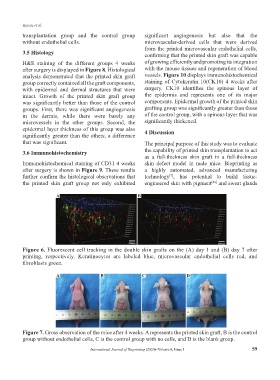
A B C D
Figure 7. Gross observation of the mice after 4 weeks. A represents the printed skin graft, B is the control
group without endothelial cells, C is the control group with no cells, and D is the blank group.
International Journal of Bioprinting (2020)–Volume 6, Issue 1 59