
Page 37 - IJB-6-3
P. 37
Shpichka, et al.
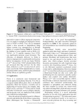
Figure 1. Tubulogenesis within native and PEGylated fibrin gels ICC – immunocytochemical staining;
L – lumen; TEM – transmission electron microscopy. Copyright permission provided by IOP Publishing .
[58]
approach to improve fibrin angiogenic properties of choice due to its good biocompatibility,
is to use single or multiple pro-angiogenic factors biodegradability, and other described biological
such as VEGF or bFGF. They can be entrapped properties in Table 3. The experience gained in
within a fiber network or immobilized using cell encapsulation was transferred and adapted to
release systems (e.g. nanoparticles) or through bioprinting.
affinity or covalent binding (Table 2). Moreover, Fibrin-based bioinks were successfully
the binding of some peptides such as RGD, LN- applied to print skin, heart, and neural constructs.
TGF-β1, or T1 peptide sequence from CCN1 Particularly, Cubo et al. fabricated a bioprinted
[87]
can significantly promote vessel formation. The skin substitute from plasma-derived fibrin and
efficiency of plasmids delivered by fibrin to primary fibroblasts and keratinocytes that were
improve angiogenesis is questionable and requires tested in vivo and showed to be similar to the
further investigation. On the one hand, Michlits native skin. Fibrin-collagen bioinks provided
et al. showed that VEGF plasmid-laden fibrin favorable environment for cells bioprinted using
[64]
gel increased skin flap survival. On the other a mobile skin bioprinting system. Hence, the
[88]
hand, Jozkowicz et al. revealed that all carrier in situ fabricated constructs accelerated wound
[83]
types used for the VEGF plasmid delivery (water, closure and re-epithelialization. Moreover, Kumar
phosphate buffer saline, and fibrin glue) stimulated et al. revealed that cardiomyocytes printed using
[89]
similar effects on capillaries growth. a fibrin-based bioink not only were viable and
3.3 Application proliferating but also expressed a specific cardiac
marker and coupled with cardiac fibroblasts.
Fibrin is a commonly used biomaterial and Wang et al. also successfully applied a fibrin-
[90]
widely applied in medicine from the 70 to 80s as based bioink to fabricate functional cardiac tissue
a surgical sealant (fibrin glue). Due to its flexible constructs that contracted synchronously and
properties, fibrin has become a versatile tool in responded to epinephrine and carbachol. Being
engineering of various tissues, for example, skin, the same complex as cardiac one, neural tissue
blood vessels, and bone. [84-86] Fibrin and its blends was achieved using fibrin which structure ensured
with other biomaterials, such as collagen, alginate, cell alignment and guided Schwann cells’ growth.
and hyaluronic acid, are applied in both scaffold Bioprinting tumors is a novel interesting
and scaffold-free technologies. direction in not only fibrin application but also
Since the advent of bioprinting and its influence in bioprinting in general. [91] The fabricated tumor
started to take hold of the field of tissue engineering, models are positioned mostly to be used as a
fibrin (fibrinogen) has become a biomaterial more relevant platform for drug screening and
International Journal of Bioprinting (2020)–Volume 6, Issue 3 33