
Page 39 - IJB-6-3
P. 39
Shpichka, et al.
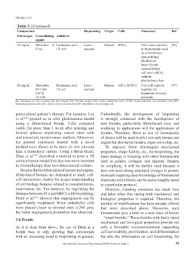
Table 3. (Continued)
Components Bioprinting Origin Cells Outcomes Ref
Fibrinogen Cross-linking Additives
agents
20 mg/ml Thrombin 4 Hyaluronic acid Laser- Human iPSCs Cells were sensitive [96]
U/ml 1% w/v assisted to biomaterials used
as a bioink base
(not printing)
Hyaluronic-
based blends
ensured better
cell survivability
without
pluripotency loss
20 mg/ml Thrombin Hyaluronic acid Laser- Human ASCs, ECFCs Cell-cell contacts [97]
40 U/ml 1% w/v assisted regulate the
CaCl2 formation of vessel
50 mM networks
Ref: References; n/a: Not available; RB: Rose bengal; PVA: Polyvinyl alcohol; iPSC: Induced pluripotent cells; HUVEC: Human umbilical vein endothelial cells; MSC:
Mesenchymal stromal cells; ASC: Adipose-derived stromal cells; ECFC: Endothelial colony-forming cells
personalized patient’s therapy. For instance, Lee Undoubtedly, the development of bioprinting
et al. [92] printed an in vitro glioblastoma model is strongly connected with the development of
using a fibrin-based bioink. Cells remained new bioinks, particularly fibrin-based ones; and
viable for more than 1 week after printing and widening its applications will the applications of
formed spheres expressing cancer stem cells bioinks. Therefore, fibrin as one of biomaterials
and metastatic invasiveness markers. Moreover, of choice will be used to print not only tissues and
the printed constructs treated with a novel organs but also tumor models, organ-on-a-chip, etc.
method were shown to be more in vivo relevant To improve fibrin (fibrinogen) mechanical
than a monolayer culture. Using a fibrin blend, properties, shape fidelity, etc., for bioprinting, the
Zhao et al. [32] described a method to print a 3D main strategy is blending with other biomaterials
cervical tumor model that also was more resistant such as gelatin, collagen, and alginate. Despite
to chemotherapy than two-dimensional culture. its simplicity, it will be further used because it
Despite the biofabrication of tissues and organs, does not need strong structural changes in protein
fibrin-based bioinks are demanded to study cell- molecule requiring deep knowledge of biomaterial
cell interactions, mainly for deeper understanding chemistry and a bioink can be easier roughly tuned
of cell biology features related to vascularization, to a particular protocol.
innervation, etc. For instance, by regulating the However, blending consumes too much time
distance between ECs and MSCs with bioprinting, and labor when fine tuning both mechanical and
Piard et al. showed that angiogenesis can be biological properties is required. Therefore, the
[93]
significantly modulated: When endothelial cells number of modifications has been already offered
were placed closer to stromal ones (≤200 μm), that were described above. Moreover, such
the better angiogenesis promotion was observed. biomaterials give a birth to a new class of bioink
3.4 Trends – “smart bioinks.” These bioinks with finely tuned
mechanical and biological properties provide not
As it is clear from above, the use of fibrin as a only a favorable microenvironment supporting
bioink base is only growing that corresponds cell survivability, proliferation, and differentiation
with an increasing trend to bioprinting in general. but also the information on cell functioning, for
International Journal of Bioprinting (2020)–Volume 6, Issue 3 35