
Page 106 - IJB-6-4
P. 106
Triple-layered coaxial nozzle for 3D bioprinting
experiments were conducted with three different and the three of them fell within the material’s
nozzles. As shown in Table 1, nozzles 1 and 2 allow printing window, as they allowed controlled and
the fabrication of single-layered hollow tubular continuous deposition of a filament. An alginate-
structures of equal outer diameter (OD), but with based bioink embedded with MG-63 cells was
different layer thicknesses. Likewise, nozzle 3 chosen for this evaluation since alginate is a widely
allows the fabrication of structures with a greater used biocompatible material, easily extrudable
diameter and layer thickness than nozzles 1 and 2. and features rapid crosslinking upon exposition
These nozzles were subsequently 3D printed with to divalent cations, which enables excellent shape
biocompatible photopolymer resins (Figure 1C) fidelity in bioprinted constructs .
[29]
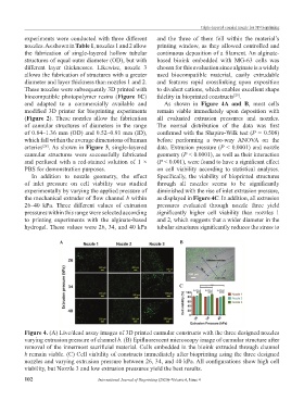
and adapted to a commercially available and As shown in Figure 4A and B, most cells
modified 3D printer for bioprinting experiments remain viable immediately upon deposition with
(Figure 2). These nozzles allow the fabrication all evaluated extrusion pressures and nozzles.
of cannular structures of diameters in the range The normal distribution of the data was first
of 0.84–1.36 mm (OD) and 0.52–0.91 mm (ID), confirmed with the Shapiro-Wilk test (P = 0.508)
which fall within the average dimensions of human before performing a two-way ANOVA on the
arteries . As shown in Figure 3, single-layered data. Extrusion pressure (P < 0.0001) and nozzle
[28]
cannular structures were successfully fabricated geometry (P < 0.0001), as well as their interaction
and perfused with a red-stained solution of 1 × (P < 0.001), were found to have a significant effect
PBS for demonstration purposes. on cell viability according to statistical analyses.
In addition to nozzle geometry, the effect Specifically, the viability of bioprinted structures
of inlet pressure on cell viability was studied through all nozzles seems to be significantly
experimentally by varying the applied pressure of diminished with the rise of inlet extrusion pressure,
the mechanical extruder of flow channel b within as displayed in Figure 4C. In addition, all extrusion
26–40 kPa. Three different values of extrusion pressures evaluated through nozzle three yield
pressures within this range were selected according significantly higher cell viability than nozzles 1
to printing experiments with the alginate-based and 2, which suggests that a wider diameter in the
hydrogel. These values were 26, 34, and 40 kPa tubular structures significantly reduces the stress to
A B
C
Figure 4. (A) Live/dead assay images of 3D printed cannular constructs with the three designed nozzles
varying extrusion pressure of channel b. (B) Epifluorescent microscopy image of cannular structure after
removal of the innermost sacrificial material. Cells embedded in the bioink extruded through channel
b remain viable. (C) Cell viability of constructs immediately after bioprinting using the three designed
nozzles and varying extrusion pressure between 26, 34, and 40 kPa. All configurations show high cell
viability, but Nozzle 3 and low extrusion pressures yield the best results.
102 International Journal of Bioprinting (2020)–Volume 6, Issue 4